 1/1
1/1
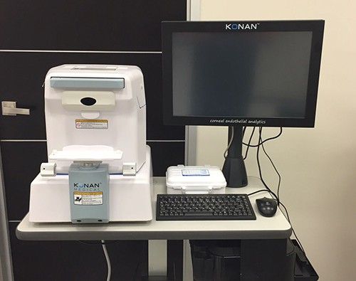
Konan CellChek XL Specular Microscope
AMERICA North (USA-Canada-Mexico)
Konan sets the standard for specular microscopy with strong clinical evidence, ease of use, and patented analysis methods that can reliably assess even problematic endothelium.
CellChek Specular Microscope
Cornea Fundamentals
Clinical Resources
Testimonials
Specifications
Support FAQs
Customer Service
Fully Automated
Konan’s CellChek specular microscope features auto-align, auto-focus, auto-caputure, auto-analysis, and auto-pachymetry for one-button ease of use. But this is where others end.
Konan additionally includes semi-automated methods (see Analysis Methods section) to make robust use of minim numbers of observable cells with advanced disease state corneas.
Trends Analysis – Only from Konan
Konan’s patented capture method acquires data samples that include position data which can allow accurate re-assessment of same specular data sample areas to trend cellular statistics over time.
Trends analysis is critical to understanding your chosen treatment responses and progression or arrest of disease.
FDA 510(k) cleared under “Specular Microscopes” Product Code NQE
Why accept anything less than the assurance of the FDA’s review for both imaging and assessment of the corneal endothelial cell layer, morphology of endothelial cells, and corneal pachymetry.