
Esaote G Scan MRI
AMERICA North (USA-Canada-Mexico)
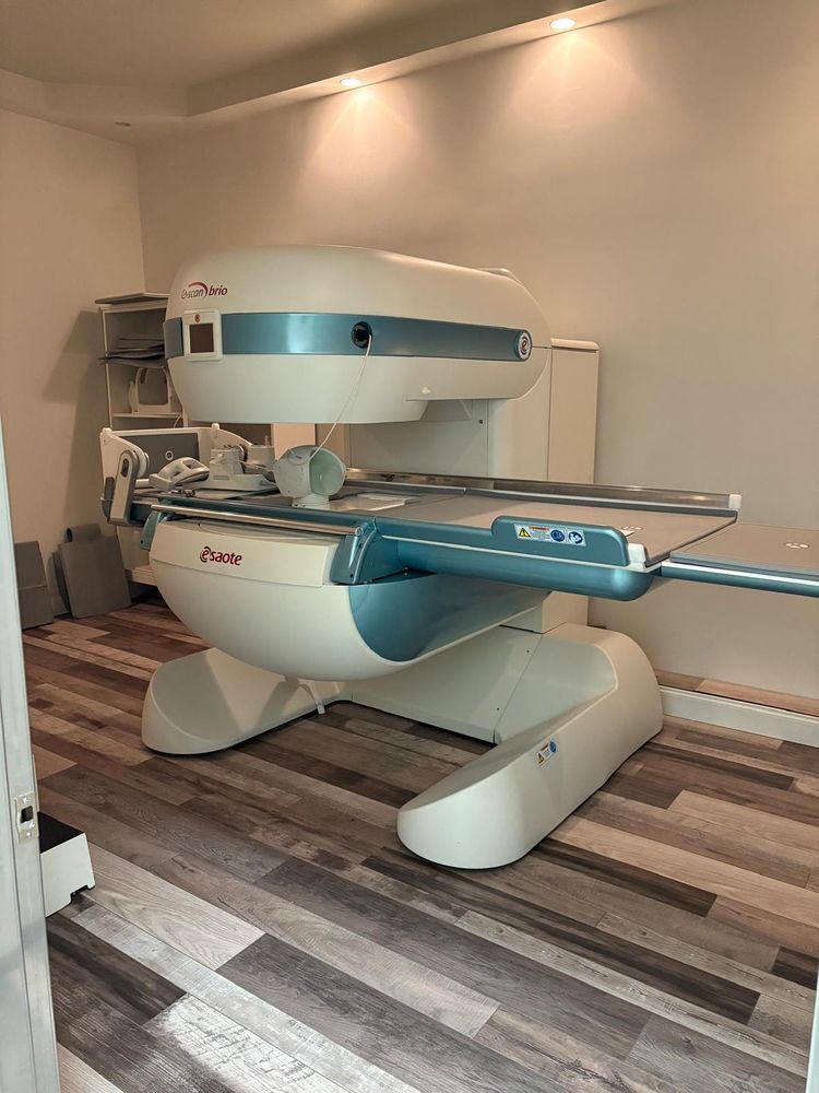
G-SCAN Dedicated MRI Unit
The G-scan magnetic resonance system is a dedicated musculoskeletal scanner,
specifically designed for limbs, joints and spine examinations.
The unique design of the magnet ensures high quality exams and
patient comfort during weight bearing or recumbent MRI exams.
Due to the compact system design, the system sites in as little as 245 square feet.
Thanks to its low energy consumption, the system can be connected to
a standard power outlet (220V).
G-SCAN Magnet
The G-Scan magnet represents the latest advances in lightweight, rare earth materials
and advanced design. It is based on years of research and design work at
Esaote coupled with customer feedback from previous magnetic resonance systems.
The unit includes:
C-shaped permanent magnet in neodymium with .25 Tesla field strength with
unique tilting capability from 0 degrees to 90 degrees in 2 degree increments
Vertical field for higher SNR and improved image quality
Generous opening of 16” to accommodate the largest patients
Fast gradient coils with ±20 mT/m gradient strength, 0.8 ms rise time
(from 0 to +20mT/m) SR:56mT/m/ms
Radio frequency transmission coil
Internal thermostatic control system
Real Time Positioning Module with LCD for instant feedback on patient positioning
and anatomy of interest
Patient table
Swivel patient table with large wide tabletop ensures maximum patient comfort.
The table feature 3 fixed location points for easy positioning of RF coils and
flexible patient positioning. Includes dedicated set of positioning cushions.
Maximum patient weight: 450lbs.
G-Scan RF System
The S-scan RF system features a highly uniform and powerful RF system capable of
meeting the demands of a busy MRI practice. It has two 1800-watt RF amplifiers and
passive receive only coils, which expedite patient positioning and
imaging parameter flexibility.
G-Scan RF Coils
Set of 9 high-sensitivity, self-centering receive coils with automatic recognition;
most coils are equipped with the Dual Phased Array (DPA) technology,
using the advanced multi-receiver phased array techniques for optimal performance.
Coils also feature built in preamplifiers and low noise electronics ensuring
excellent image quality.
All coils come with a set of dedicated padding cushions, for patient comfort and
to reduce patient motion.
DPA Shoulder coil
Advanced shoulder imaging with ergonomic design for comfort.
DPA delivers highest SNR and image quality. (16.8 x 15.4 x 7.9 cm).
Shoulder coil
Solenoidal coil for maximum positioning flexibility, designed especially for
large shoulders and knees (14.5 x 17.5 x 12.6 cm).
DPA Knee coil
DPA coil for comprehensive views of the knee designed for
patient comfort and acceptance and large coverage area. (14.3 x 16.0 x 18.3 cm)
DPA Hand-Wrist coil
DPA coil designed for comfortable, high resolution imaging of the wrist and
hand. (11.9 x 7.2 x 20.0 cm)
DPA Foot and Ankle coil
DPA coil designed for optimized and natural positioning of the foot and
ankle and elbow (14.5 x 10.0 x 28.5 cm).
Flexible multi purpose coil
The flexible coil is a general-purpose coil. It is used when positioning with
the standard coils is difficult due to patient limitations. (Painful joint, inability to
assume a certain position). Includes Velcro positioning strap. (4 x 34 x 28 cm).
G-scan Standard Spine package
The G-scan spine package consists of spine coils for optimized imaging of
the cervical and lumbar spine.
X-Large DPA Lumbar Spine coil
The lumbar spine coil package features advanced coil design combining a flat coil built in
to the coil base and a removable belt coil which encompasses the patient.
There are 1 belt, X-large and to guarantee optimum imaging of the lumbar spine
regardless of patient habitus.
Cervical Spine coil
The c-spine coil is designed for easy patient positioning while delivering excellent images
of the cervical spine. It has a unique integrated flexion-extension mechanism for
imaging different neck positions.
Included in the package are a set of cushions and bolsters for stable and
comfortable positioning of the patient during the exam.
G-scan Advanced Spine Package
The G-scan Advanced Spine Package consists of 3 spine coils providing higher
signal uniformity and increased signal-to-noise.
4 channel Lumbar Spine Coils-Qty 2
The coils feature a 20% increase in SNR and two different sizes (medium and large)
to accommodate a larger patient population. The coils can be used for hip imaging
providing a 40% increase in SNR over the previous flex coil option. Included are a
set of cushions and bolsters for stable and comfortable positioning of
the patient during the exam.
Dual Phased Array Cervical Spine Coil
The new DPA C-spine coil provides better coverage signal uniformity and a 20% increase
in SNR. The coil sits into the patient bed and includes positioning cushions for
patient comfort.
Imaging parameters R 4.1 EVO Package
The G-scan has been designed to provide high quality imaging of the spine and
extremities using standard and advanced MRI pulse sequences.
Patient imaging times and throughput are optimized.
Scoutview: real time orthogonal images displayed on LCD panel
Scan planes: axial, sagittal, coronal, oblique and double oblique
Minimum slice thickness: 2mm in 2D .6 mm in 3D
Image matrix: 128 to 512 x 512
Maximum number of slices: 256
Saturation slabs for artifact reduction with graphic positioning
Multi-Pack and Multi angle
Flow compensation
Pulse sequences:
multiplanar scout
spin echo T1 (SET1)
spin echo T2 (SET2, SET2 SA)
multi echo (SE_PD_T2)
inversion recovery (IR)
short TI inversion recovery (STIR, STIR T2 S, STIR T2 A)
spin echo half echo (SET1HE)
spin echo half scan (SET1HF)
turbo SE T2 (TSE, TSE S, TSE SA, TSE SP)
turbo ME (TME)
gradient echo (GE, GE T2, GE T2 FC)
short time inversion recovery gradient echo (GE-STIR)
gradient echo 3D (T3D_T1)
speed spin echo T2 #3 (SSE-SET2 #3)
o FSE (Fast Spin Echo T2 Weighted)
speed spin echo T2 (SSE-SET2, SSE-SET2 S, SSE-SET2 SA,
SSE-SET2 SP)
speed spin echo T2 #1 (SSE-SET2 #1)
speed spin echo T2 #2 (SSE-SET2 #2)
real time positioning sequence
2D and 3D HYCE (maximizes contrast between CSF, spinal cord and nerve roots)
fast FLAIR
XBone (Dixon Fat/Water separation)
FSE STIR (fat suppression for spine exams)
3D steady State (3D SHARC) SS T1, SS T2
MAR (Metal Artifact Reduction)
2D and 3D HYCE
STIR T2 weighted
FSE PD/T2 (cuts imaging time by 40%)
SPEED-UP (reduces 2D sequence time by up to 30%)
MAR (Metal Artifact Reduction)
True Motion: (2D HYCE real-time kinematic sequence)
G-scan eXP-EVO Package
The G-scan eXP package has been designed to improve the performance of the scanner
by enhancements to acquisition speed and image quality via new hardware and
software.
Enhancements include:
-Advanced GPU/CPU Image Reconstruction
-2D and 3D Speed Up Technology
-Metal Artifact Reduction
short time inversion recovery gradient echo (GE-STIR)
gradient echo 3D (T3D_T1)
speed spin echo T2 #3 (SSE-SET2 #3)
o FSE (Fast Spin Echo T2 Weighted)
speed spin echo T2 (SSE-SET2, SSE-SET2 S, SSE-SET2 SA,
SSE-SET2 SP)
speed spin echo T2 #1 (SSE-SET2 #1)
speed spin echo T2 #2 (SSE-SET2 #2)
real time positioning sequence
2D and 3D HYCE (maximizes contrast between CSF, spinal cord and nerve roots)
fast FLAIR
XBone (Dixon Fat/Water separation)
FSE STIR (fat suppression for spine exams)
3D steady State (3D SHARC) SS T1, SS T2
MAR (Metal Artifact Reduction)
2D and 3D HYCE
STIR T2 weighted
FSE PD/T2 (cuts imaging time by 40%)
SPEED-UP (reduces 2D sequence time by up to 30%)
MAR (Metal Artifact Reduction)
True Motion: (2D HYCE real-time kinematic sequence)
G-scan eXP-EVO Package
The G-scan eXP package has been designed to improve the performance of
the scanner by enhancements to acquisition speed and image quality via
new hardware and software.
Enhancements include:
-Advanced GPU/CPU Image Reconstruction
-2D and 3D Speed Up Technology
-Metal Artifact Reduction