 1/1
1/1
GE
AMERICA North (USA-Canada-Mexico)
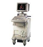
GE LOGIQ 3
GE Healthcare’s LOGIQ 3 is an advanced ultrasound instrument so versatile and flexible that it can be used in a full range of clinical applications from OB/GYN, vascular and cardiac to neonatal studies. The LOGIQ 3 offers the most economical, compact and portable design – making it the ideal choice for office-based practices or wherever economical full-function ultrasound scanning is required
Features
TruScan Architecture
Fifteen-inch high-resolution monitor
Integrated mode and gain controls help ensure fast and efficient studies.
Simple, intuitive single-hand operation.
Two active probe ports for ease of use
Small system footprint (19.7” w X 37.2” d)
Highly-maneuverable, four-wheel platform with front-wheel lock
Ultra-mobile, easily moves from room to room
Ergonomic user interface, with standard, full-featured ultrasound keyboard, increases user comfort and productivity
Automatic Optimization to improve image quality and consistency
Easy3D integrated for improved diagnostic confidence from viewing images in other planes
Virtual convex creates a convex field of view for a 30% increase in image size in linear transducers
Comprehensive reporting packages matched to your needs
Flexible on-board image management
B-mode gain, dynamic range and gray scale maps to bring out subtle detail
Color invert, color maps and on/off capability
Doppler gain, baseline shift, sweep speed, angle adjust and spectral invert for Doppler optimization after scanning
Reconstruct a stored cine loop into a 3D image for virtual rescanning
Measurements without calibration
System Specifications
Height: 1360 mm (53.4 in)
Width: 502 mm (19.7 in)
Depth: 948 mm (37.2 in)
Weight: approx. 153 kg (337 lb)
Monitor: 15-inch Non-Interlace, High-Resolution, and Flat Display
Applications
Abdominal
Obstetrical
Gynecological
Cardiac
Musculoskeletal
Vascular
Urological
Small Parts and Superficial
Pediatric and Neonatal
Transcranial
Transducer Types
Sector Phased Array
Convex Array
Microconvex Array
Linear Array
Operating Modes
B-Mode (2D)
M-Mode (M)
Anatomical M-Mode
Color Flow Mode (CFM)
Power Doppler Imaging (PDI) with Directional Map
PW Doppler with High PRF (PW)
M-Color Flow Mode
Steerable CW Doppler (TBD)
Dedicated CW Doppler (TBD)
Transducers
GE 3.5C – 2-5MHz, Convex
GE 3C – 2-5MHz, Convex
GE 5C – 3-9MHz, Convex
GE E8C – 4-11MHz, Microconvex
GE 3S – 1.5-3.5MHz, Sector Phased Array
GE 7S – 3-9MHz, Sector Phased Array
GE 10LB – 5-10MHz, Linear