 1/1
1/1
GE
AMERICA North (USA-Canada-Mexico)
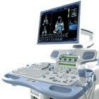
GE Vivid 7 Dimension
Features
Advanced Tissue Synchronization Imaging (TSI) provides quantification of LV synchronicity information. Time-to-peak data derived from TSI’s simple-to-use “red-light / green-light” visualizations let you quantify left ventricle synchronicity for heart failure patients or those undergoing cardiac resynchronization therapy (CRT).
Automated Function Imaging (AFT) is an easy-to-use tool for assessing and quantifying left ventricle wall motion. AFI (derived from 2D strain) delivers reproducible, systolic, quantitative segmental and global assessments of the left ventricle – quickly and accurately.
Real-Time 4D Color Flow Full Volume – Assess hemodynamic information in color in real time in the same heart cycle – with the option of viewing a 6-slice cross section of the left ventricle.
Contrast Imaging enhances visualization of the LV border for wall motion analysis and ejection fraction calculations in multidimensional imaging and 4D full volume imaging
6 Slice – can further enhance full volume color information. The 6-slice view takes the full volume color information and creates 6 equidistant slices and the ability to focus on a specific region of interest.
Multiple-angle compound imaging enhances border definition, reduces acoustic artifact and improves contrast resolution.
The 9L transducer features a new ergonomic profile, with a smaller, more ergonomic handle design and a smaller, lighter, more flexible cable, for easier, more comfortable scanning.
Speckle Reduction Imaging – offers new improvements that enhance image quality in difficult-to-image patients.
Blood Flow Imaging (BFI) gives you a better understanding and delineation of directional blood flow in vessels.
IMT measuring technology dramatically reduces the time it takes to measure the carotid artery’s intima-media thickness, which studies show may be an early sign of athersclerosis.
Multi-Dimensional and 4D Imaging – with multi-dimensional imaging, you can simultaneously acquire bi-plane and tri-plane images from the same heartbeat – without moving the probe’s position – to get more cardiovascular information in less steps.
9 Slice allows you to visualize regional wall motion in a whole new way by slicing the left ventricle of a 4D full-volume dataset into nine equidistant short-axis views.
4D Coded Phase Inversion (CPI) enhances your contrast imaging capabilities on your 3V transducer, allowing you to get excellent sensitivity, resolution and tissue suppression in 4D imaging.
Flexible reporting package increases productivity by allowing you to choose the report configurations, and edit the findings text and conclusion sections according to your lab’s individual workflow needs.
Fast-pull down menus help you automatically sort and insert information.
Customizable normal report values let you define measurement values.
MPEGVue allows you to export more compressed studies.
EVUE simplifies PC setup for remote interactive image monitoring.
DICOM connectivity, with embedded raw-data storage, permits post-exam quantitative analysis.
System Specifications
Height (with 21” LCD monitor): 54.1 in (137.5 cm)
Width: 25.2 in (64.0 cm)
Depth: 35.4 in (90.0 cm)
Weight (with LCD monitor, without peripherals): 403 lbs (183 kg)
Display Screen: 21-inch High-Resolution (up to 1600 x 1280 pixels), flicker-free LCD monitor
Applications
Cardiac
Vascular
Transducers Types
Linear Array
Phased Array
Convex Array
Microconvex Array
Volume ‘4D’ Phased Array
Multiplane TEE
Pencil Doppler
Operating Modes
B-Mode (2D)
M-Mode
B-Flow (BF)
Color Doppler
Color M-Mode
Spectral Doppler
CW Doppler
PW Doppler w/ high PRF
Anatomical M-Mode
Transducers
GE 3V – 1.5-4.0MHz, Volume ‘4D’ Phased Array
GE M4S – 1.5-4.3MHz, Phased Array
GE 3S – 1.5-3.8MHz, Phased Array
GE 5S – 2.2-5.0MHz, Phased Array
GE 7S – 2.9-8.0MHz, Phased Array
GE 10S – 3.7-11.5MHz, Phased Array
GE 9L – 3-10MHz, Linear
GE 10L – 4-10MHz, Linear
GE 12L – 4.9-13.0MHz, Linear
GE M12L – 4.9-13.0MHz, Linear
GE i13L – 5.3-14.0MHz, Linear (Intraoperative)
GE i8L – 3.9-10.0MHz, Linear (Intraoperative)
GE 4C – 1.6-5.0MHz, Convex
GE 5C – 3.0-6.7MHz, Convex
GE 8C – 3.7-8.0MHz, Convex
GE M7C – 2.9-7.0MHz, Convex
GE E8C – 3.7-8.0MHz, Microconvex (Endocavity)
GE 6T – 2.7-7.0MHz, Multiplane TEE (Transesophageal)
GE 9T – 3.1-10.0MHz, Multiplane TEE (Transesophageal)
GE P2D – 2.0MHz, Non-Imaging CW Doppler Pencil
GE P6D – 5.0MHz, Non-Imaging CW Doppler Pencil