 1/1
1/1
GE
AMERICA North (USA-Canada-Mexico)
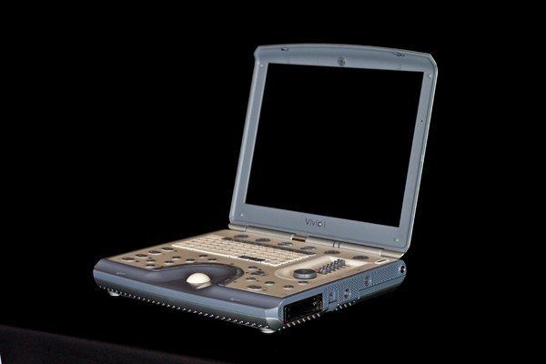
GE Vivid i
Features
Full diagnostic and monitoring capabilities
Advanced measurement and analysis tools
New information management tools
Phased-array transducer technology for 2D, color and Doppler imaging.
Multiple focal zones help optimize image quality.
Extremely high frame rates, enhanced color flow and color angio assistant in acquiring extremely low- velocity flows.
5 levels of Coded Octave Harmonics help you obtain quality images from difficult-to-image patients.
LVO contrast option, based on 2D Harmonic imaging with Coded Phase Inversion, aids in scanning your most challenging patients.
Triplex and duplex display capabilities to simplify Doppler acquisitions.
Automatic Tissue Optimization (ATO) option helps you obtain quality images faster by automatically adjusting the image settings to optimize images.
Complete measurement and analysis worksheets and reports are tailored to each exam.
Live Anatomical M-Mode corrects for off-axis orientation in situations where the heart is not positioned or shaped normally.
Tissue Doppler Imaging provides real-time Doppler spectral information for specified myocardial motion, allowing for instantaneous tissue velocity measurements.
Over 40 gigabyte disk space for archive storage
Full DICOM connectivity with embedded raw data speeds exams by allowing you to perform post-exam quantitative analyses at your convenience on the integrated EchoPAC or optional EchoPAC Dimension workstation.
MPEGvue option compresses images into compact file sizes. These high quality images can be transferred to any compatible media or network, or e-mailed directly for viewing.
eVue option permits remote interactive monitoring of images on any PC, via wired or wireless network communication, for fast and convenient consultations.
Lightweight, 5 kg package makes system transport fast and easy – simply strap on your back or carry to your patients.
Ultra-small footprint reduces operational costs by eliminating the need for special rooms, lifts or vans.
Rechargeable battery provides up to 1.0 hour of full scan operation.
System Specifications
Height: 59 mm (2.3 in)
Width: 358 mm (14.2 in)
Depth: 313 mm (12.4 in)
Weight: approximately 5 kg (11 lbs) without battery
Display Screen: 15-inch High-Resolution, TFT LCD
Applications
Cardiac
Vascular
Abdominal
Small Parts
Transducer Types
Linear Array
Convex Array
Phased Array
Doppler Pencil
Multiplane TEE
Operating Modes
B-Mode (2D)
M-Mode (M)
Color Doppler
Power Doppler Imaging (PD)
PW Doppler with high PRF (PW)
CW Doppler (CW)
Tissue Doppler Imaging (TD)
M-Color-Mode (MC)
Color Flow Mode (C)
Transducers
GE 3S-RS – 1.5-3.6MHz, Phased Array
GE 5S-RS – 2-5MHz, Phased Array
GE 6S-RS – 2.7-8.0MHz, Phased Array
GE 7S-RS – 3.5-8.0MHz, Phased Array
GE 10S-RS – 4.5-11.5MHz, Phased Array
GE 8L-RS – 4-13MHz, Linear
GE 12L-RS – 6-13MHz, Linear
GE i12L-RS – 5-13MHz, Linear
GE 4C-RS – 1.8-6.0MHz, Convex
GE 8C-RS – 4.7-11.0MHz, Convex
GE P2D-RS – 2.0MHz, Doppler Pencil
GE P6D-RS – 6.0MHz, Doppler Pencil
GE 6T-RS – 2.9-8.0MHz, Multiplane TEE
GE 9T-RS – 4-10MHz, Multiplane TEE