 1/1
1/1
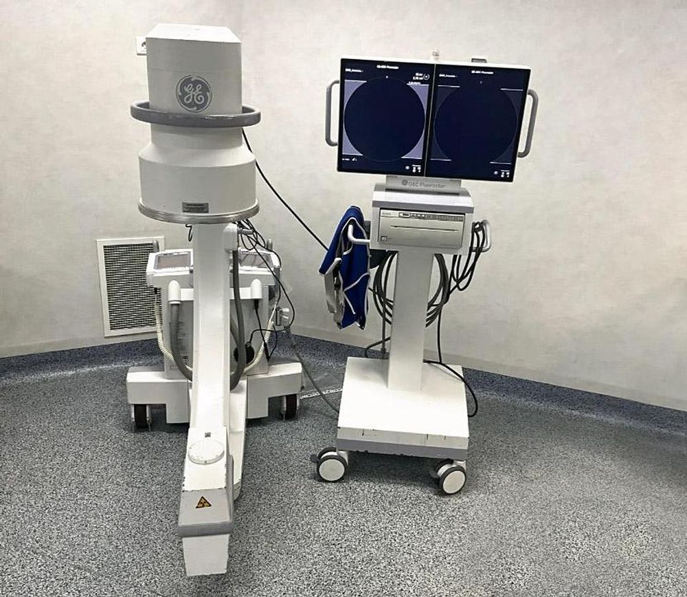
General Electric OEC Fluorostar
EUROPE (Western and Northern)
Composed of :
• A rolling module (Fluorostar MC) with 2 OEC FLUOROSTAR screens +
brightness amplifier (OEC 09TO)
• A Sony UP-970AD hybrid graphic printer
• Software version REV04.00.06
• Display versions: 4.00.58, BV: 4.00.33
• Installed options: Bitmap, Dicom
Features :
Generator
• High frequency 20 kHz
• 2.2 kW Monobloc
• Up to 110 kVp
• Up to 8 mA in fluoroscopic mode
• Up to 20 mA for exposing radiographic films
• High-performance pulsed mode up to 8 mA
• Pulse width: 50 ms
• Digital spotlight up to 8 mA
X-ray tube
• Fixed anode X-ray tube
• Nominal focal points of 0.5 and 1.5 mm
• Total filtration of the entire tube assembly > 3.9 mm Al
Collimator Preview
• Display of the collimator position on the screen
• Iris PreView and dual-blade collimator
• Independently adjustable asymmetric shutters
• Adjustment of collimators without X-ray exposure
Standard fluoroscopic mode
• kVp range: 36-110
• mA range: 0.2-3
• Automatic and manual fluoroscopic modes
Fluoroscopic hip mode
• kVp range: 36-110
• mA range: 0.2-5.4
• Automatic and manual fluoroscopic modes
Low-dose fluoroscopic mode
• kVp range: 36-110
• mA range: 0.2-3
• Automatic and manual fluoroscopic modes
Pulsed fluoroscopic mode
• kVp range: 36-110
• mA range: 0.2-5.4
• Pulse frequency: 1, 2, 4, 8 Hz
• Automatic and manual fluoroscopic modes
Low-dose pulsed fluoroscopic mode
• kVp range: 36-110
• mA range: 0.2-3
• Pulse frequency: 1, 2, 4, 8 Hz pps
• Automatic and manual fluoroscopy modes
High-performance pulse mode
Fluorescent mode
• kVp range: 36 to 110
• mA range: up to 8
• Pulse rate: 1, 2, 4, 8 ips
• Automatic and manual fluoro modes
• Pulse duration: 50 ms
Digital spot mode
• kVp range: 36 to 110
• 8 mA
• Automatic exposure stop
• Automatic image recording
Radiographic mode
• mA range: 20
• mAs range: up to 80
• Exposure time: 0.1 to 4 seconds
• Computer-controlled exposure time
• Cassette holder: 25.4 cm x 30.5 cm
Video Imaging System
Precision imaging features
• Automatic window: Automatically adjusts brightness and contrast to produce a high-quality image.
• Automatic control of exposure rate
• Average filter
• Edge enhancement filter
• Predefined imaging profiles: standard, dense anatomy (hip),
hand, thorax, reduced dose.
23 cm Image Intensifier
• Tri-mode image intensifier tube 23 cm/15 cm/11 cm
• Minimum central resolution (on monitor):
- 9" (23 cm): 2.0 lp/mm
- 6" (15 cm): 2.8 lp/mm
- 4.5" (11 cm): 3.2 lp/mm
• DQE: 65% (typical)
• Grid: 8:1 ratio
Video camera
• High-resolution CCD camera 1000 × 1000 pixels
• Fully digital interface
• Fixed gain control
Video display
• 19" (48 cm) monochrome TFT-LCD flat screen
- Tilt: 23 cm
- Viewing angle: 160° horizontal and vertical
• Minimum brightness: 800 cd/m²
• Maximum brightness: 1000 cd/m²
• Minimum contrast ratio: 800:1
• High resolution 1280 x 1024
• External video output:
- DVI-I connector (compatible with VGA and DVI-D analog signals)
- Fixed resolution of 1280 x 1024 pixels
Image processing
• 1K x 1K x 12-bit image processing
• Dynamic recursive filter with motion adaptation
- Allows the user to adjust the noise filter levels for optimal image quality
- Guarantees optimal image quality even in case of movement
• Automatic control of digital brightness and contrast
• Manual control of digital brightness and contrast
• Improved contours
• Image negation mode
• Automatic image recording and saving
• Image exchange
• Last image retention (LIH)
• Zoom (up to 1600%) and image navigation
• Left-right inversion
• Up-down inversion
• Digital image rotation
- Real-time 360° rotation for dynamic and static images
- Image positioning without additional exposure
• Image annotation
- Laterality (left/right)
- Comments
• Storage for 15,000 images
Exam management
• Patient information
- List of exams
- Personalized patient information
• Display and summary of the DAP/X-ray dose
• Export and print the DAP/X-ray dose summary
• DICOM interface (optional)
- Storage
- Impression
- Work list
- Structured Radiation Dose Report (RDSR)
- MPPS