 1/1
1/1
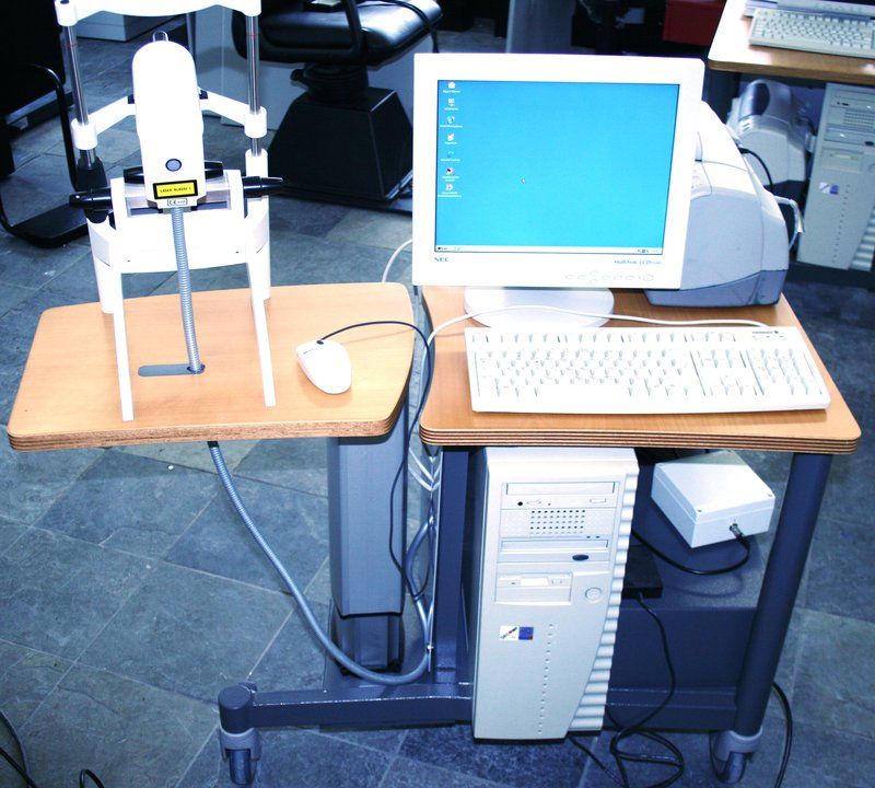
Heidelberg HRT II Retina Tomograph
EUROPE (Western and Northern)
Used, good tested and maintained condition
1 year Schairer house guarantee
Scope of delivery:
Heidelberg Engineering HRT II Retina Tomograph, electrical orig. Lifting table
Printer
PC, monitor, keyboard, mouse
Foot pedal
Operating instructions German
HRT Retina module
Macular diagnostics at a glance
Simple, early detection of edematous changes
Three-dimensional representation of the retina thickness
Precise detection of changes through image alignment with TruTrack™
No data interpolation, therefore lower risk of overlooking pathological changes
Show and Tell function to educate the patient
Modular, expandable platform
Network-capable
In a steadily aging population,
the number of cases of age-related macular degeneration
and diabetic retinopathy continues to increase.
The HRT Retina Module offers a quick,
straightforward method for assessing the macula
without data interpolation.
Retinal edema index
The edema index makes it easy and quick
to locate suspicious areas of the retina
that are not readily apparent to doctors,
even during a thorough examination
with a contact lens. HRT depicts changes
in the retina that occur due to small fluid deposits.
These changes are often not classified
as clinically significant macular edema,
but may be an indicator of an increased risk of the disease.
Based on this information, a decision can be made
as to whether the patient should be examined more frequently.
Using the retinal thickness measurement values,
the HRT Retina Module creates a complete map
of the recorded area without interpolating data.
Retinal thickness maps may be more useful
than the edema index for monitoring the progression
of pathological changes.
In addition, the three-dimensional maps can be used
to explain retinal abnormalities to the patient more clearly.
The reflection image is another HRT tool used
to detect anomalies. It shows differences
in the appearance of the retinal surface
and allows evaluation of the edema index
and retinal thickness maps.
Technical data
Device dimensions
Workstation: 1075 x 516 mm
Device type:
Confocal scanning laser ophthalmoscope
Field of view:
15° x 15° (transversal)
Scanning depth:
1.0 to 4.0 mm (automatic)
Optical resolution:
10 µm per image point (transversal) x 62 µm per pixel (longitudinal)
Reproducibility:
Height measurement ± 20 µm
Size raster image:
3-D images: up to 384 x 384 x 64 pixels
Image file size:
4-6 MB (compressed) per eye
Recording time per 3D image:
1-6 seconds
Refraction range:
-12 to +12 diopters spherical, -6 to +6 Diopters cylindrical
Pupil diameter:
≥ 1 mm
Wavelength:
670 nm
Transportability:
Optional notebook version and carrying case
Viewer software:
Glaucoma, retina or cornea
Glaucoma modules:
Moorfields regression analysis (MRA)
Retinal nerve fiber layer thickness (RNFL) analysis
C/D Ratio
Retina Analysis
Glaucoma Probability Value (GPS)
Ethnically-specified databases
OU symmetry analysis
9-point quality control
Parameters adapted to the size of the optic nerve head
Direct findings analysis
Topographical course analysis (progression)
Retina Modules:
Edema index
Retinal thickness
3-D thickness map of the retina
Flexible positioning of the ETDRS Grid
trend analysis
cornea modules:
400×400 µm image view
2 µm intervals, up to 80 automatic cross sections
1 µm resolution
Semi-automatic endothelial cell counting
Sequence scan, cross section scan, volume scan.
HRT Glaucoma Module
A reliable tool for predicting changes
Reliable prediction of structural changes proven by numerous studies
Progression analysis has been validated with patient data from over 10 years
Optic disc analysis more meaningful than expert assessment
Extensive databases selectable by ethnicity
Analysis of asymmetries
Expandable platform
Network-capable
The Heidelberg Retina Tomograph (HRT)
is the gold standard for detecting glaucomatous changes
in the optic nerve head (papilla)
and on the peripapillary nerve fiber layer
as well as for monitoring the progression
of progressive glaucoma in everyday clinical practice.
It offers very special support for the early detection
of structural changes in everyday life,
before a visual field restriction can be measured.
The excellent diagnostic performance of HRT
is widely recognized. It is based on fast,
non-invasive, reproducible topography measurements,
which are automatically converted into high-resolution
two- or three-dimensional images by the HRT software
without data interpolation.
The image acquisition without prior pupil dilation does
not restrict the patient, so that he can even drive
on the road without any problems after the measurement.
At the same time, live fundus images
and automatic quality control during
and after recording support the user
and ensure consistently high image quality.
The previous study on the Ocular Hypertension
Treatment Study (OHTS) showed that the analysis
of optic disc changes led to the early detection
of incipient glaucoma in 55% of all cases,
even before detectable visual impairment occurred.
HRT has been shown to be a reliable tool
for predicting glaucoma.
The OHTS also shows that HRT detected damage
to the temporal-superior sector in 40% of patients,
which later developed into overt glaucoma,
based solely on the initial examination.
The diagnosis was later confirmed during
an average 5-year follow-up.
Equally impressive was the fact that 93%
of the patients in whom HRT did not detect any pathological changes during the initial examination had not yet developed glaucoma after 5 years.
Of all available diagnostic imaging devices used to diagnose and treat glaucoma, the HRT glaucoma module offers the highest image resolution. It provides a quantitative assessment of all relevant anatomical features, such as: B. the excavation shape of the optic nerve head, the neuroretinal rim volume and the peripapillary retinal nerve fiber layer. When analyzing these characteristics, both the individual optic disc size and the patient's age are taken into account.
Moorfields regression analysis (MRA) was developed at Moorfields Eye Hospital in London. MRA differentiates between glaucomatous and healthy optic discs by detecting and evaluating diffuse and focal changes in the neuroretinal rim. The method takes into account optic disc size, which plays an important role in assessing the neuroretinal rim, as well as age-related changes. The MRA classifies the eye based on standard values and gives the results for the entire optic disc and for six individual sectors separately. The results are presented as color-coded symbols: a green check mark represents -within normal limits-, a yellow exclamation mark represents -borderline- and a red cross represents -out of normal limits. The substudy of the Ocular Hypertension Treatment Study (OHTS) recognized MRA as highly predictive of the likelihood of developing open-angle glaucoma.
Glaucoma is a progressive disease in which it is crucial to document and quantify changes or the progression of the disease over time in order to confirm the diagnosis, improve patient care and adapt treatment to the individual.
HRT topographic change analysis (TCA) is one of the best-known and most valued analysis methods in glaucoma diagnostics. Early diagnosis and documentation of changes in the optic nerve head and the retinal nerve fiber layer is of fundamental importance for progression analysis. The TCA analyzes repeatable changes over time for topography and altitude variations and displays these in red. The greater the change, the more intense the color.
The HRT combines high speed with efficient TruTrack software and is therefore unsurpassed in its performance and clinical benefit. The TruTrack software works in the background and tracks over 500 points on each image to precisely map follow-up scans to the images from the initial examination and reliably detect even small changes.