 1/1
1/1
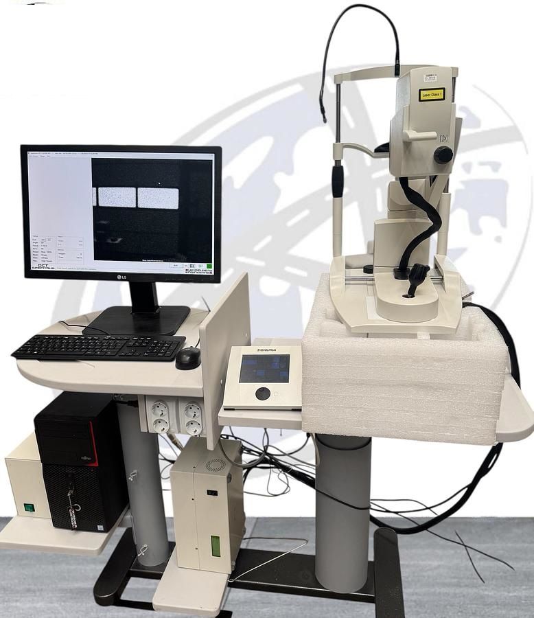
Heidelberg Spectralis
EUROPE (Western and Northern)
IR
Infrared Reflectance Imaging
Uses an 820 nm infrared laser.
Great for general fundus imaging and patient eye tracking.
Also used as a base for aligning OCT or other scans.
BAF
Blue Autofluorescence (BluePeak)
Uses 488 nm blue laser to detect autofluorescence from
lipofuscin in the RPE.
Helps detect early retinal pigment epithelium (RPE) damage
(e.g., AMD, inherited retinal diseases).
BAF + IR
Combined Blue Autofluorescence and Infrared Imaging
Simultaneously captures both BAF and IR images.
Useful for comparing structural and functional layers of
the retina in one scan.
Grid Icon (Blue Dot Pattern)
Scan Area or Pattern Selector
Lets you choose specific scan regions (e.g., macula, optic disc).
Commonly used for setting raster patterns, volume scans,
or focal point targeting.
H S
(Icon with arrow over horizontal bars – just partially visible)
Likely “HS” = High-Speed or High-Resolution Scan Mode Toggle
Lets you switch between:
HS (High Speed) = Faster scan, lower resolution.
HR (High Resolution) = Slower scan, higher detail (especially useful in OCT).