 1/1
1/1
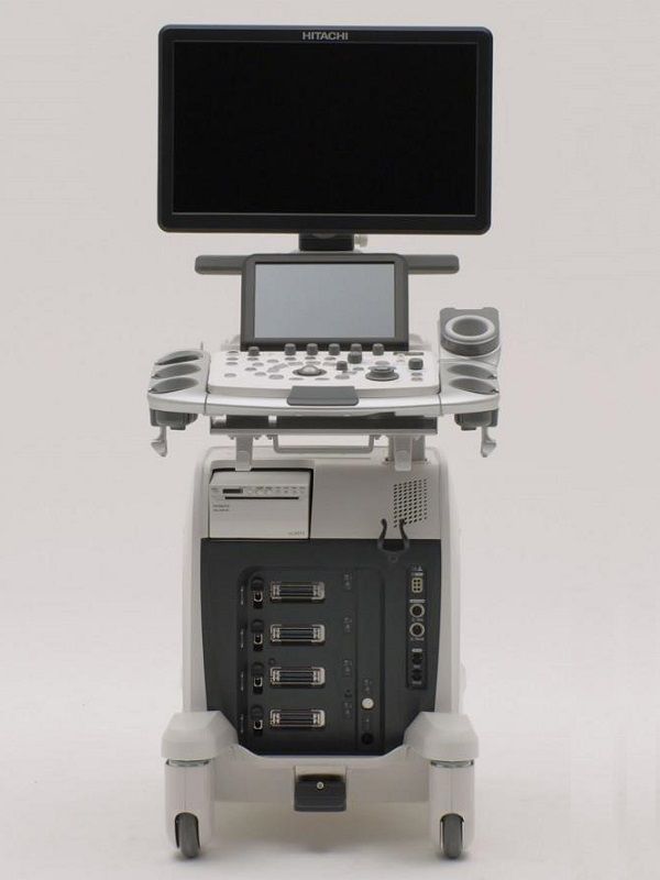
Hitachi Arietta 65
EUROPE (Western and Northern)
Hitachi ARIETTA 65 – Diagnostic Ultrasound System
Expertly designed to optimize productivity
ARIETTA 65 Ultrasound has been designed to perform quick and
precise diagnosis in general imaging without compromising on
productivity and workflow.
This ultrasound platform combines productivity, enhanced tools and
technology to provide
Smooth workflow and productivity
Superb imaging and accurate diagnosis
Simple to use applications and streamlined practice
ARIETTA 65 further offers:
21.5’’ LCD widescreen monitor, optimal for quick and detailed observation
Touch panel, positioned at an easy-to-operate angle
Operator console designed for ease of use
Protocol Assistant, features that allow to custom previously registered protocols and
automatically prepare the next step in the exam, prompt you through the exam by
reducing keystrokes and preventing duplications or omissions.
Intelligent Automatic functions for smooth workflow
Auto Optimizer: Automatically adjusts the gain and baseline position and velocity range
with just one button
Cardiac Features: automation tools enable faster and
smoother cardiovascular examination, building on data acquired by
our premium systems
Mobility that meets autonomy – allowing to scan approx. 1 hour in battery mode for
smooth use in emergency care or changing rooms
Power cord hook, handles and folding mechanism, allowing to move the system safely
Superb Imaging, powered by top-performing imaging technologies
Top-performing imaging technologies have been mitigated for
enhanced diagnostic confidence and precision, namely:
High definition dynamic Tissue Harmonic Imaging (HdTHI) resulting in
superb spatial resolution and penetration
High Resolution Imaging (HI REZ) to emphasize tissue structures and
enhance contrast resolution for greater clarity
Compound Imaging for clear visualization of tissue boundaries, high contrast resolution
and speckle reduction to allow clearer observation of lesions
Simple to use applications, enhancing precision in a wide user range
ARIETTA 65 incorporates advanced tools for diverse clinical use and
detailed evaluation:
Real-time Tissue Elastography(RTE)
Assesses tissue strain in real time and differences in tissue stiffness as a color map –
useful in a wide variety of clinical fields such as breast, thyroid gland and
urinary structures.
Estimates liver fibrosis staging in patients with hepatitis C (LF Index) with
the abdominal convex transducer
Supports time-saving automation features to improve objectivity,
reproducibility and productivity
Contrast Harmonic Imaging (CHI) – provides homogeneous enhancement throughout
the field of view to enhance diagnostic capability.
Panoramic View – generates a single, elongated image by moving the probe across the
target organ, thus allowing a larger field of view and enhanced diagnostic precision.
Interventional support tools
Needle Emphasis – automatically adjusts the deflection angle of beams and
images to enhance needle visibility and assist in safe and accurate punctures.
Marking Assist to support marking on the body surface before surgical operation.
Automatic tools for vascular and cardiac
Auto IMT
Automatically measures the max and mean values of Intima-Media Thickness (IMT) for
reproducible and accurate follow up of vascular disease
Dual Gate Doppler
Collects Doppler waveforms from two locations during the same heart beat
Enables faster and more accurate measurement of LV diastolic performance indicators
Useful in diagnosis of fetal arrhythmia and Carotid stenosis
2D Tissue Tracking (2DTT)
Speckle tracking technique that quantifies the movement of the entire left ventricle or
the local movement of the myocardium resulting in dynamic analysis of myocardium.
ADDITIONAL INFORMATION
WEIGHT 85 kg
DIMENSIONS 53,5 × 74 × 126,5 cm
MEDICAL SPECIALTY
Cardiology, Endocrinology, Gastroenterology, Neurosurgery, Obstetrics – Gynecology,
Radiology, Rheumatology, Surgery, Urology, Veterinary
COLOR Color Flow Mode, eFLOW
DOPPLER CW Color Doppler, Dual Gate Doppler, PW Color Doppler,
Real-time Doppler Auto Trace
DISPLAY TECHNOLOGY LCD Monitor
IMAGE QUALITY WXGA (1600 x 900)
SCREEN DIMENSIONS 21.5″ inches
TOUCH PANEL 10.1″ inches
CONNECTIVITY Analog Video Input/Output, DICOM, DVI-D, Ethernet, USB 2.0, DVD External
PRINT TYPE Ethernet Color Printer, Thermal B/W Printer, USB Color Printer
HEAD PORTS Electronic scanning probes: 4 active
SYSTEM PORTABILITY Wheeled
COMPATIBLE PROBES
Convex
Hitachi C35 Abdominal Convex
Hitachi C251 Abdominal Convex
Hitachi C253 Abdominal Convex
Hitachi C22P Biopsy Convex
Hitachi C22K Intraoperative Convex
Hitachi C25P Biopsy Convex
Hitachi C41 Convex Pediatric
Hitachi C42 Convex Pediatric
Hitachi C42K Intraoperative Convex
Hitachi C42T Intraoperative Convex
Endocavity
Hitachi C41V Endocavity Vaginal
Hitachi C41V1 Endocavity Vaginal
Hitachi C41B Endocavity Vaginal/Rectal
Hitachi C41RP Endocavity Rectal
Linear
Hitachi L34 Vascular Linear
Hitachi L441 Vascular Linear
Hitachi L442 Vascular Linear
Hitachi L55 Breast Linear
Hitachi L64 Vascular Linear
Hitachi L43K Intraoperative Robotic
Hitachi L44K Intraoperative Linear
Hitachi L53K Intraoperative Linear
Hitachi L44LA Intraoperative Linear
Hitachi L51K Intraoperative Robotic
Hitachi L46K1 Intraoperative Linear
Phased Aray
Hitachi S211 Phased Array
Hitachi S11 Phased Array
Hitachi S31 Pediatric Phased Array
Hitachi S3ESEL TEE Cardio
RT-3D (4D)
Hitachi VC35 Convex 3D/4D
Hitachi VC41V Vaginal 3D/4D
Bi-Plane
Hitachi CC41R Endocavity Bi-Plane
Hitachi CC41R1 Endocavity Bi-Plane
Hitachi CL4416R Endocavity Bi-Plane
Hitachi C41L47RP Endocavity Bi-Plane
Radial
Hitachi R41R Endocavity Electronic Radial
Independent CW Doppler
Aloka UST-2265-2 Independent CW Doppler