 1/1
1/1
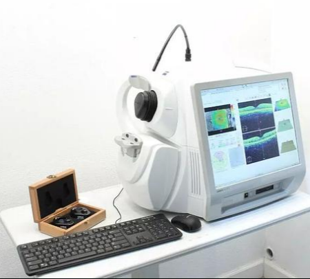
Humphrey Zeiss Cirrus 500 OCT
AMERICA North (USA-Canada-Mexico)
The Humphrey Zeiss Cirrus 500 OCT Tomographer
with Windows 10 (lenses not included).
This unit comes fully refurbished by an in house trained
and certified technician. Each unit is inspected mechanically,
optically, and even cosmetically with FREE lifetime phone support
and a 6 month parts and service warranty.
The Zeiss Cirrus 500 HD-OCT is a Spectral domain
optical coherence tomography (SD-OCT) device for
comprehensive eye care, providing essential high-resolution retinal
and optic nerve imaging for glaucoma and retinal disease management,
plus anterior segment imaging for corneal issues, all in an easy-to-use,
cost-effective unit with smart features like automatic alignment and
data registration to streamline workflows and track disease progression.
Key Features & Capabilities:
Diagnostic Imaging: Delivers sharp, detailed cross-sectional images
of the retina (Macular Cube, Radial Line) and optic nerve (RNFL analysis).
Glaucoma & Retinal Analysis: Includes tools like RNFL/GCC analysis,
Macular Thickness Analysis, and Guided Progression Analysis (GPA) to
monitor changes over time.
Anterior Segment Imaging: Offers high-resolution scans of
the cornea and anterior chamber (with optional modules)
for corneal disease assessment.
Ease of Use: Features an intuitive interface, automated alignment
(Auto Fovea Finder, Auto Registration), and the ability to
scan through small pupils (even 2mm) without dilation.
Workflow Efficiency: Uses smart features like Auto Repeat and
Auto Registration to maintain consistency between visits,
improving patient flow.
Advanced Technology: Employs Selective Pixel Profiling and
Enhanced Depth Imaging for optimal image quality,
even in challenging cases.
Clinical Applications:
Managing Glaucoma: Assessing optic nerve damage and
tracking progression.
Diagnosing Retinal Diseases: Analyzing macula health and
detecting subtle changes.
Cataract Surgery: Assessing the retina before surgery.
Evaluating Corneal Conditions: Imaging the cornea
and anterior chamber.