
 1/1
1/1
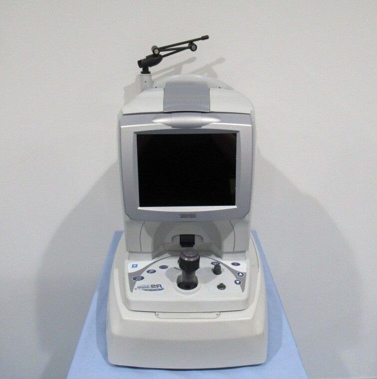
Nidek RS‑3000 Advance OCT
MIDDLE EAST
The NIDEK RS-3000 Advance Optical Coherence Tomography (OCT) system is a
high-performance imaging platform designed for advanced retinal diagnostics,
glaucoma assessment, and anterior segment analysis.
Utilizing spectral domain OCT technology, it delivers ultra-high resolution images,
rapid scan speeds, and precise layer segmentation for accurate clinical evaluation.
With integrated confocal scanning laser ophthalmoscope (SLO) fundus imaging,
en face OCT capability, and powerful analysis software,
the RS-3000 Advance supports comprehensive eye care from screening to
complex pathology management. Optional anterior segment modules expand
its utility to corneal and angle measurements, making it an ideal solution for
ophthalmology clinics seeking speed, accuracy, and reliability.
This unit is available for sale and in excellent condition,
ready to enhance diagnostic capabilities.
Spectral Domain OCT with optical resolution: Z: 7 µm, X-Y: 20 µm; digital resolution: Z: 4 µm, X-Y: 3 µm.
High Scan Speed – Up to 53,000 A-scans/sec for faster exams and reduced motion artifacts.
Scan Range – X: 3–12 mm (12 mm for line scan), Y: 3–9 mm, Z: 2.1 mm.
Light Source – Superluminescent diode (SLD) at 880 nm.
Fast 3D Acquisition – 1.6 seconds in regular mode.
Integrated Fixation – Internal (637 nm) and external (630/565 nm) fixation lamps.
Auto Alignment with Z-direction adjustment.
Small Pupil Imaging – Minimum pupil diameter: 2.5 mm.
Focus Range – -15 D to +10 D.
Working Distance – 35.5 mm.
Advanced Retinal Analysis – Segmentation of 6+1 retinal layers, macular thickness map,
RNFL thickness map, NFL+GCL+IPL analysis, optic nerve analysis, and follow-up analysis.
Confocal SLO Imaging – 785 nm source, 40° × 30° field of view (zoom to 20° × 15°).
Tiltable 8.4″ Color LCD Display for ease of use.
PC Networking – Compatible with NAVIS-EX image filing software with DICOM connectivity.
En Face OCT – Visualization of retinal pathology and vascular structure with selectable depth offsets.
Image Averaging – Up to 120 images for high clarity.
Optional Anterior Segment Module – Corneal thickness measurement/map,
anterior chamber angle (ACA) measurement, AOD500/750, and TISA500/750.
Flexible Scan Patterns – Retina: macula line, cross, map, multi, radial, disc circle,
map, radial. Cornea: line, cross, radial, ACA line.
Follow-up and Tracing Functions – Torsion eye tracker, Tracing HD Plus, HD checker,
select & rescan, auto shot.
Compact Dimensions – Main unit: 380 (W) × 524 (D) × 499–531 (H) mm; weight 34 kg.