 1/1
1/1
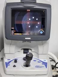
Nidek RS-3000 Lite
EUROPE (Western and Northern)
Used equipment inspected by our experts
Functional condition: very good
Cosmetic condition: very good
Nidek reliability and precision
Sold with computer
Technical specifications
OCT for general screening
The RS-3000 Lite offers optimal cost-performance thanks to its
retinal surface imaging system. It was designed for screening in
general ophthalmology consultations.
Retinal surface imaging by phase-shift OCT
The RS-3000 Lite uses a different method than phase-shift OCT to
image the retinal surface, replacing SLO.
Wide scan (9 x 9 mm)
The wide 9 x 9 mm scan area allows for analysis of the [NFL+GCL+IPL] layers at
and around the macula, around the optic disc, and even in the periphery.
*The [NFL+GCL+IPL] layers are composed of the nerve fiber layer (NFL),
the ganglion cell layer (GCL), and the inner plexiform layer (IPL).
High-speed scanning (53,000 A-scans/s max.)
High-speed scanning (53,000 A-scans/s max.) and high-speed averaging (50 images max.)
produce a high-definition B-scan image.
Multifunctional monitoring
Multifunctional monitoring allows for the analysis of all data obtained by OCT
and detailed observation of chromosomal changes in retinal thickness and condition.
This function displays the progression of the pathology over time.
The comparison mode displays two user-selected images.
Displaying progress
Displaying comparison
Custom report
Report layout can be customized, and data from separate reports for each scan
model can be summarized into a single report to avoid printing multiple pages.
Comparison between the RS-3000 Advance 2 and the RS-3000 Lite
Model RS-3000 Advance 2 RS-3000 Lite
Receiver Surface Imaging SLO (12 frames per second)
40° x 30° Field of View Fundus OCT (1.8 fps)
36° x 30° Field of View
Scanning Speed Up to 85,000 scans A/s Up to 53,000 scans A/s
OCT Sensitivity Normal, Fine, Ultrafine Normal, Fine
Normative Database Area 9 mm x 9 mm (macula)
6 mm x 6 mm (disc) ←
Normative Database Long Axial Length 9 mm x 9 mm (macula) ←
OCT Angiography (in (Option) Available / Not available
Scanning pattern (retina) Macular line (scan angle adjustable in 1° increments)
Macular cross (with cross scan / without cross scan)
Macular map
Multi-macula (X-Y: 5 x 5)
Radial macula (6 lines / 12 lines)
Disc circle
Disc map
Radial disc (6 lines / 12 lines) Macular line (scan angle adjustable in 15° increments)
Macular map
Multi-macula (X-Y: 5 x 5)
Disc map
Scanning pattern (cornea) with
optional anterior segment module Corneal line
Corneal cross
Corneal radial (6 lines / 12 lines)
ACA line Corneal radial (6 lines / 12 lines)
ACA line
Average images Max. 120 images Max. 50 images
Choroidal mode Available Not available
Torsion eye tracer Available Not available
Trace tracking Available Not available
Trace analysis Available ←
HD trace plus Available Not available
HD checker Available Not available
Flexible cross scan Available Not available
Selection and re-scan mode Available Not available
Auto-shoot (for capturing follow-up images) Available Not available
Internal fixation target Cross shape (laser) Circular shape (LED)
21-inch PC screen 17-inch screen
Features
OCT for general screening
Fundus phase OCT for imaging the fundus surface
Wide scan (9 x 9 mm)
High-speed scan (max. 53,000 scans A/s)
Multifunctional tracking
Custom report