 1/1
1/1
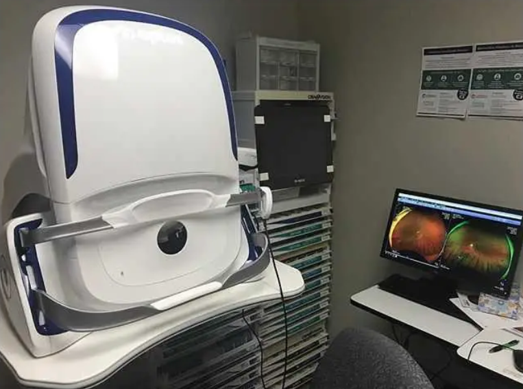
Optos Monaco Ultra WideField Retina Imaging
ASIA (South East)
Image Modalities
Color
Red-free
Choroidal
Autofluorescence
OCT
Image Views
Standard: 200⁰ Single Capture
Auto-montage: Up to 220⁰
Central Pole: Detailed view of the macula
Stereo: Image pairing for optic disc and retinal evaluation
OCT: Cross-sectional imaging of ocular structures, including the fundus
Monaco offers the following benefits:
• UWF with integrated OCT saves time, space and minimizes patient movement
• Central pole OCT provides comprehensive multi-modal imaging
• optomap images and OCT scans are correlated to facilitate pathology examination
• Color, AF, and OCT images are shown in a single, comprehensive view
Optos Monaco Features and Benefits
• UWF with integrated OCT saves time, space and minimizes patient movement.
• High resolution 200º single-capture optomap images improve pathology detection and management frommacula through the far periphery.
• Non-mydriatic, cSlO imaging through most cataracts and small pupils (2 mm).
• 3-in-1 Color Depth ImagingTM provides important clinical data from the retinal surface through the choroid.
• Green laser autofluorescence minimizes patient exposure to blue light and shows macula and optic nerve head detail.
• Central pole OCT provides comprehensive multi-modal imaging.
• optomap images and OCT scans are correlated to facilitate pathology examination.
• Color, AF, and OCT images are shown in a single, comprehensive view.
• Fast, comfortable image acquisition is easier on patients and improves clinic flow.
• OptosAdvance Image Management facilitates image review and consultations and includes measurement and auto montage capabilities.
Technical Specifications
Image Modalities
optomap color and optomap plus (red and green laser):
Color composite view
Green laser view
Red laser view
optomap af (green laser): autofluorescence
Optical Coherence Tomography (OCT)
Resolution
optomap color: 20 μm
optomap plus, af : 14 μm
Wavelengths
Red laser: 635 nm
green laser: 532 nm (for af )
Exposure Time
Less than 0.4 seconds
Tomographic Imaging
Signal Type: Optical scattering from tissue
Signal Source: Super luminescent Diode (SlD) 830 nm
Optical Power: laser safety Class-1 following IEC/en60825-1:2014
Typical Axial Resolution: