 1/1
1/1
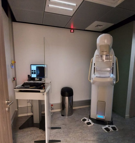
Philips Microdose L30
RUSSIA and Central Asia
No scattered radiation or electronic noise:
The image is formed by scanning with a patented multi-slit collimator,
which filters out 97% of scattered radiation.
Individual photon signals are clearly separated from background electronic noise.
This significantly reduces image noise, allowing for highly efficient dose utilization.
No data loss during signal conversion: Direct registration of
individual X-ray quanta occurs without analog-to-digital conversion,
so there is no data loss associated with such conversion.
No ghost images:
Since the Philips detector has a high readout speed, allowing it to
register every incoming photon, there are no ghost images that could interfere with
image interpretation or slow down imaging.
50-µm pixel size:
The small pixel size allows for visualization of fine structures in the breast,
such as microcalcifications and spicules, at half the radiation dose.
100% fill factor:
Thanks to the design of the new photon-counting detector, 100% of its surface is active,
allowing all photons that hit it to be recorded.