 1/1
1/1
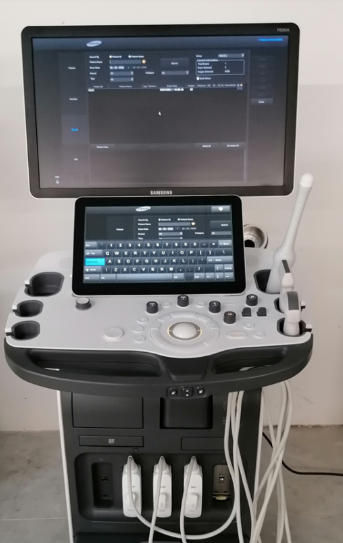
Samsung RS80A
RUSSIA and Central Asia
The RS80A is a state-of-the-art premium ultrasound scanner manufactured
by Samsung Medison. In 2010, Samsung and Medison merged,
enabling them to produce high-quality and reliable products for
the most challenging clinical situations. Samsung Madison specializes in
the development of medical devices,
and its ultrasound equipment boasts high accuracy and safety.
Stunning visualization, high frame rates, a wide range of transducers,
and original innovations solve medical problems of any complexity.
This device has proven itself in hospitals and clinics where the
highest standards for medical devices and
high-quality patient care are paramount.
This expert-class scanner features a 23-inch high-resolution LED
graphic display and a touchscreen panel for easy and
comfortable operation. Up to five sensors can be installed,
plus one additional port, as well as a flash drive slot, a DVD-RW drive,
and intelligent recording of examination fragments with
additional editing and rewind options make this device one of
the most cost-effective acquisitions for your business.
Doctors value high-quality images of organs and systems for
accurate examination and subsequent diagnostics, to avoid missing
pathologies and detect them in the early stages.
Therefore, Samsung Medison has developed its professional ultrasound
equipment with superior image quality and detail across a variety of
diagnostic areas.
Possibilities
Clear Vision delivers crisp, clear photos with reduced grain,
enhanced edges, and increased contrast. Improves your photos in real time.
Multi Vision – reduces artifacts and increases detail,
optimizes insonation angles;
Sono View – data can be archived and viewed later, involving other doctors
in working on the patient, provides access to copying to
a digital video disc and flash drive (USB).
Visualization
B – 2D study using the gray scale, THI;
M – mode (heart), and with the cardio package it is possible to perform
1-dimensional examination of the heart, anatomical M-mode,
color M-mode;
Dopplers: CD, PD, DPDI, TDI, PW, CW, HPRF;
3D – volumetric sensors in static mode and gray scale scan 3D, the structure
of the vessels is restored in CD and PD modes;
4D is the latest technology that allows for real-time viewing; thanks to
volumetric sensors, 3D scanning occurs (with the Live 3D package for
observing structures and functions);
It is possible to divide the interface into several areas: up to two, four or
more images are displayed on the monitor, as well as in B/C, B/PD modes in
real time;
Built-in mixed modes: B/M, B/PWD, B/C, B/PD, B/PD/PWD, B/C/PWD;
Linear sensors have a trapezoid mode;
Zoom, which provides the most detailed viewing of images, which will help
to better assess the current state of the organs being examined;
The presence of an ECG module will help to avoid using
many additional research products;
Additional feature: viewing panorama;
HDVI – visualizes the image and makes it three-dimensional,
increasing the clarity of tissue with different echo density
(using this function, it is possible to diagnose various injuries and
pathological changes in the object);
Realistic Vue is an option that helps reconstruct 3D ultrasound images
(close to real-life images) using artificially created and superimposed light.
A special algorithm can reproduce 3D fetal anatomy with
a high level of detail.
Natural Vue is a unique, highly accurate, realistic image reconstruction
feature that will help you perform virtual amnioscopy and
various morphological studies for many structures in 3D mode
(with Realistic Vue).
STIC – dynamic volumetric visualization of the fetal heart;
AutoIMT – calculates the intima-media thickness. This is an important feature for assessing the early development of atherosclerosis, stroke, and myocardial infarction.
Elastoscan – elastography for examining the thyroid gland, mammary gland
and prostate gland to determine tissue stiffness and softness;
E-Strain – selection of two zones for comparison and
quantitative assessment of tissue elasticity (if Elastoscan is available);
E-Thyroid – allows for comparative elastography without compression
(semi-automatic);
E-Breast module – selection of 1 zone for elastography (automatic);
S-Shearwave – liver elastography, with determination of the stiffness index
of various areas, obtaining reliable data on malignancy;
S-Detect Breast – detects and analyzes neoplasms in the mammary gland,
measures according to the standards of data results with an assessment of
the risk of tissue malignancy;
S-Detect Thyroid – detects thyroid tumors through Ti-Rads (malignancies);
S-Fusion – combines and displays magnetic sensors on
image sensors obtained by various methods – CT, MRI, ultrasound;
S-Tracking – navigation for precision insertion of the biopsy needle;
CEUS+ contrast study;
Arterial Analysis – precise analysis of the carotid artery (its elasticity and wall thickness). 3D capabilities available;
Strain+ – segmented assessment of myocardial contractility
(independent automated calculation);
ADVR – studies are recorded on a flash drive at the current moment;
A pedal used for remote control;
MSV – viewing multiple slices on a monitor (3D study);
VolumeCT – cubic display (Cube Sectional View) or 3 intersecting planes (Cross View);
OVIX – selects parts of 3D frames;
Mirror View – 3D display of structures from the front, left, right and
top – mirrored;
Multi OVIX – allows you to view several different OVIX images at
the same time.
Innovations
S-Vue sensors (monocrystalline) – piezoelectric elements made from
1 crystal;
Quick Scan – press one button for instant,
more detailed examination customization
(optimal parameters thanks to organ recognition in the smart database);
S-Harmonic – provides the highest quality visualization and is independent
of scanning depth. This is an improved tissue harmonic function;
SRF – noise reduction;
VCE – removes blurry areas, enhances the quality of images in 3D display;
VSI – 3D images show objects better using shades from 1 of
5 possible color maps;
FSI – selective contrast enhancement of images/echograms at any depth;
Inversion 3D – creation of a “cast” for the area of interest, with a
deeper assessment of vascular structures thanks to the inverse 3D option;
Color STIC – volumetric reconstruction of the heart contractions of
the fetus under study together with CD in real time;
EZ Exam – step-by-step ultrasound examination protocols and inclusion of
the necessary modes during the study process;
S-Vision – a thin beam that reduces noise, increases the clarity and depth of
ultrasound scanning;
HQ-Vision – an option for optimizing the display of anatomical structures of
the musculoskeletal system;
Needle Mate – a biopsy needle with enhanced display contrast. It also
features an additional feature for changing the ultrasound wave angle
(linear transducer, beam steering).
Packages
Using the Samsung RS80A ultrasound machine in the field of gynecology, it
is possible to scan various formations (volume) and
pathological processes with malignancy.
Obstetrics: a variety of studies related to the management of
pregnant women.
Fetal heart: measurements in B and M-mode;
Cardio: M-modes, CW, TD;
Research: carotid arteries, arteries of the upper and lower extremities, veins
of the lower extremities, vessels of the abdominal cavity;
Urology;
Abbreviations: LP/PP, MZP, MPP, ZSLZ, LV/RV, EF, PFSMR, PSS/KDS.
Conveniently select the right transducer for
efficient healthcare professionals.
The interface's work area displays the scan of interest,
trimester selection (for gynecological examinations),
the selected transducer, and the ability to change the settings later.
Connectors for up to four transducers are available.
Sensors (type BF/IPX7) for Samsung RS80A ultrasound equipment
Mini-convex sensors, linear, sector phased, intracavity micro-convex,
three-dimensional, pencil.
Contact – convex sensors, linear, sector phased, intracavity microconvex,
three-dimensional sensors.
Technical specifications
Weight 62 kg
Permissible load during operation 75 kg
Dimensions 1380x450x700
Touch screen 13.3”
LED monitor 23”
Storage capacity of snapshots Up to 8200