 1/1
1/1
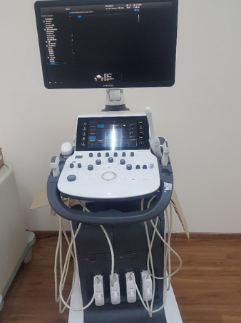
Samsung WS80A
RUSSIA and Central Asia
Unique image quality, innovative technologies and
advanced volumetric ultrasound capabilities.
The Samsung Medison ultrasound machine offers a variety of
diagnostic modes and programs, making it suitable for
a variety of ultrasound examinations. Superior image quality,
combined with the latest imaging technologies,
ensures confident medical staff.
Functions
The EVO hybrid beamformer—an advanced proprietary technology—
combines noise reduction with efficient ultrasound signal post-processing,
enabling high-frame-rate imaging in 2D, 3D, 4D, and CD modes.
This advanced feature enables the system to receive, transmit,
and process highly detailed signals, improving diagnostic performance.
S-Harmonic – with the advent of a new era of ultrasound diagnostics,
a pulse inversion option has appeared, which will provide a
high-quality ultrasound image with clear fragments without noise;
Tissue harmonic intensity (THI) is the harmonic component of
oscillations of anatomical structures when an ultrasound pulse
passes through the body;
Clear Vision – a real-time acoustic image optimization filter that
removes noise, enhances 2D image contrast, sharpens edges,
and increases contrast at the edges. This feature was developed by
Samsung, which is committed to high-quality medical diagnostics for
patients.
S-Vue transducers (CV1-8A transducer, CA1-7A transducer,
CA2-9A transducer, CA3-10A transducer) are single-crystal transducers with
a wide frequency range. Volume transducers offer high sensitivity and
image homogeneity compared to standard transducers.
Depth resolution ensures excellent image quality regardless of
the technical complexity of the situation.
The transducers are convenient and comfortable to use,
reducing fatigue for medical personnel.
The large angle of the intracavitary probe provides a wide examination field
of up to 210 degrees, visualizing a huge amount of data during scanning of
the pelvic organs (assessment of the cervix and body of the uterus, ovaries,
etc.).
5D technologies
Heart Color (Fetal Heart Study) – scanning the heart for changes in
blood flow as part of an echocardiogram (ECG) study.
The display shows standard slices (9) based on STIC and CD;
5D CNS+ (fetal brain measurements) – consists of nine axial, sagittal, and
coronal transverse views of the fetal brain, along with international brain
assessment standards and ISUOG recommendations.
This will help simplify the measurement process;
5D Limb Vol (fetal weight estimation) - quickly measures the length and
dimensions of the upper arm or femur at three points, providing an
accurate estimate of fetal weight and additional nutritional information;
5D NT (nuclear translucency measurement) – first-trimester measurements
of the nuchal translucency thickness and intracranial structures for chromosomal mutations. This unique feature accurately determines
the fetal position;
5D Follicle (follicle measurement) – size, number and volume of
follicles during a gynecological examination.
Image visualization
Crystal Vue is a cutting-edge 3D imaging technology that features
innovative intensity, gradient, and transparency settings to
enhance visualization of structures in a single image.
This technology provides physicians with superior diagnostic accuracy
and quality. The resulting image is exceptionally high quality,
improving the efficiency and reliability of medical personnel.
Crystal Vue Flow is an extension of Crystal Vue technology to
provide a deep understanding of the relationship between
anatomical structures by enhancing morphological information
with hemodynamic imaging;
Realistic Vue – realistic representation of the fruit when choosing
the direction of the light source with adjusted shadows;
CEUS+ in 3D/4D mode – gynecological assessment of
fallopian tube patency, condition of uterine tissue, endometrium, etc.
using contrast in 3D/4D;
The IOTA-ADNEX (classification of ovarian neoplasms) is a classification of
malignant ovarian tumors. It complies with the recommendations of
the IOTA group.
Multitasking WS80A
ElastoScan (including for gynecology) – a high-sensitivity mode with
ElastoScan technology helps detect pathological lesions early.
This method is easier and more informative than
conventional examinations;
S-Detect Breast – automatically delineates the contours of
the suspected breast tissue and assesses the size and characteristics of
the lesion. It is scored using the Bi-Rads scoring system.
E-Breast (ElastoScan for women's breasts) – independently calculates the
stiffness of the selected tissue and the surrounding space;
E-Thyroid (ElastoScan for the thyroid gland) is a reliable method for
assessing and documenting tissue stiffness, the option will be an
excellent addition to standard scanning. It is a highly effective method for
assessing and documenting tissue stiffness, E-Thyroid technology will
be a reliable addition to conventional gray-scale scanning,
providing clearer visualization of tumors;
E-Cervix (Cervical Elastography) E-Cervix is a tool that measures
cervical stiffness using elastography to provide additional information
useful in predicting preterm labor and successful induction of labor.