 1/1
1/1
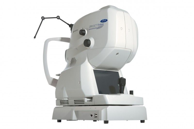
Topcon 3D Dri OCT Triton Plus - Angio
EUROPE (Central and Eastern)
Reconditioned (used),
Technical condition: very good,
Visual condition: very good,
Real product photos ,
Japanese production,
Power supply: 230 V,
Frequency: 50/60 Hz,
Power: 250 VA,
The DRI OCT TRITON PLUS is the highest-end,
most advanced multimodal model. In addition to
basic functions and FAF, it also allows for fluorescein angiography (FA),
useful in the advanced diagnosis of retinal vascular diseases.
Swept Source Technology (SS OCT) offers new possibilities in
imaging ocular structures. Compared to the most common
spectral imaging devices (SOCT), it enables significantly
higher scanning speeds, much better penetration of
ocular structures down to the sclera, and better final image resolution.
The SS OCT TRITON's 1050nm wavelength (infrared)
provides significantly better tissue penetration than
the red light used in SOCT. Infrared light also undergoes less scattering,
allowing for better visualization of the posterior vitreous cortex and
its structure. Thanks to the features described above,
the swept source DRI OCT TRITON is a device that allows for
a deeper look into the structures of the human eye.
The device incorporates extensive glaucoma functions
with a normative database. Thanks to its large scanning area,
it is possible to perform imaging and qualitative diagnostics of
the macula, measure the thickness of the nerve fiber layer around
the disc, ganglion cells in the central area,
and obtain topographic data of the optic disc in a single scan.
Main features:
simultaneous acquisition of high-quality images of the vitreous body,
retina, choroid and sclera, visualization of the vitreous humor
with the possibility of showing the differentiation of its structure,
automatic creation of maps of the thickness and
volume of the retinal and choroid layers,
obtaining significantly better quality OCT scans in
patients with reduced transparency of optical media,
including cataracts and unfavorable refractive error,
thanks to the large maximum scan width (12mm),
the device can simultaneously image the macula and nerve disc area,
high light transmittance and low light scattering allows for
obtaining high-quality, artifact-free cross-sections of
the anterior segment,
no scanning lines visible to the patient, which often cause loss
of fixation and deterioration of the examination quality,
Specification:
Observation and photography of the fundus of the eye
Photo type: Color, red-free,
Photography angle: 450, Equivalent to 300 (digital zoom),
Working distance: 34.8 mm,
Minimum pupil diameter:
Normal: 4.0 mm or more,
Narrow: 3.3 mm or more,
Observation and photography of the fundus tomogram
Scanning range (fundus of the eye):
Horizontal: 3 to 12mm,
Vertical: 3 to 12mm,
Scan profiles:
3D scan,
Linear scan (linear, cross, radial),
Scanning speed: 100,000 A-scans per second,
Horizontal resolution: 20 μm,
Axial resolution:
Digital: 2.6 μm,
Optical function: 8 μm,
Minimum pupil diameter: 2.5 mm or more,
Observation and photography of the fundus / fundus tomogram
Fixation point:
Internal fixation point:
Organic EL point matrix,
The display position can be changed and adjusted,
The display method can be changed,
Peripheral fixation point:
It is displayed according to the position of the internal fixation point,
External fixation point,
Observation and photography of the anterior segment
Photo type: IR,
Working distance: 17 mm,
Observation and photography of an anterior segment tomogram
Working distance: 17 mm,
Scanning range (on the cornea):
Horizontal: 3 to 16mm,
Vertical: 3 to 16mm,
Scan profiles:
3D scan,
Linear scan (linear, radial),
Scanning speed: 100,000 A-scans per second,
Fixation point:
Internal fixation point,
External fixation point,
TOPCON IMAGEnet6 software version 1.03 (1.35.20386), in English with lifetime license,
Keyboard on the control panel in: English,
The whole thing is placed on a mobile table OPTOPOL SE-275 (has traces of use) with electric height adjustment in the range of: 64-84 cm,
Included in the set:
User manual in PL, EN,
PC with WIN8 EN + keyboard, mouse,
LG 23” IPS 23MB35PYI monitor,
HP Color LaserJet CP1215 Printer,
TOPCON Kit AA-1 anterior segment examination attachment,
Fixation point,
Case,
Cable set,
Dimensions:
OCT: 50 x 28 x 60 cm,
Table top: 120 x 52 cm,
Overall: 52 x 140 x 140 cm,
Libra:
Table 72 kg,
OCT: 22 kg,
Overall: 135 kg,
Has a valid Technical Passport issued