 1/1
1/1
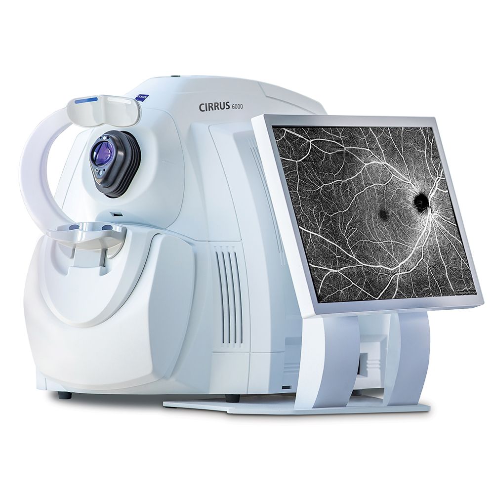
ZEISS Cirrus 6000
AMERICA North (USA-Canada-Mexico)
270% faster OCT scans and 43% faster OCTA scans
OCT cube scans in as little as 0.4 seconds
High-speed imaging in combination with FastTrac eye tracking technology reduces the chance of motion artifacts such as those caused by blinks and saccades
Greater detail: View more in seconds and dig deeper with high-definition imaging.
12×12 mm single-shot OCTA cube scan in addition to 8×8, 6×6 and 3×3 mm scans
High-Definition AngioPlex scans (8×8 and 6×6 mm) for even greater microvascular
2.9 mm scan depth
Performance OCT — faster, wider, with a new level of detail
ZEISS CIRRUS 6000 empowers clinicians with a larger field of view in a single scan,
and captures high-definition OCT/OCTA scans that reveal finer details of the retinal microvasculature −
all of which provides more insight into the patient’s condition in less time.
Proven analytics:
CIRRUS-powered treatment decisions
As the pioneering OCT technology, the CIRRUS platform offers clinicians extensive,
clinically-validated applications for retina, glaucoma and anterior segment.
The result:
precise analysis, faster throughput and smarter decision-making across a wide spectrum of clinical conditions and patient types.
Retina
Macular Change Analysis The CIRRUS data cube automatically stores and delivers each patient’s historical data to provide a variety of change assessments,
including macular thickness change maps that help you understand your patient‘s response to treatment.
Because every CIRRUS cube is tracked and registered to OCT scans from prior visits using CIRRUS’ FastTrac™ Retinal Tracking Technology, you can confidently measure point-to-point changes in macular thickness.
AngioPlex Metrix OCTA Quantification
AngioPlex® Metrix™ for Macula and ONH:
AngioPlex Metrix allows clinicians to objectively assess and track progressive eye diseases such as diabetic retinopathy
and glaucoma with quantification tools such as Vessel Density, Perfusion Density,
and Foveal Avascular Zone (FAZ) for the macula, and Capillary Flux Index for the optic nerve head. *Example In Pictures*
Glaucoma
The CIRRUS suite of glaucoma analysis tools are designed to help you better visualize, detect, and manage all stages of glaucoma,
from glaucoma suspects and mild glaucoma to severe glaucoma.
CIRRUS RNFL thickness deviation maps have been shown to be superior for detecting localized RNFL defects, compared to traditional peripapillary RNFL thickness measurements
Ganglion Cell Analysis helps identify macular glaucomatous damage, which can be missed with RNFL analysis alone
Combined GCL/IPL and RNFL thickness deviation maps provide a comprehensive widefield assessment
AutoCenter™ – ZEISS’ patented algorithm automatically identifies the optic nerve head using Bruch’s Membrane Opening (BMO) in 3-dimensions for more precise measurement of the neuro-retinal rim,
accounting for tilted discs, disruptions to the RPE and other challenging pathology
Unique to ZEISS, Guided Progression Analysis™ (GPA™) provides both trend and event-based analyses that detect statistically significant change and quantify rate of change for key RNFL, ONH, and GCL/IPL parameters
Anterior Segment
Anterior Segment Premier Module CIRRUS also enables comprehensive imaging and quantification of the anterior segment for refractive surgery planning and follow-up, corneal evaluation and glaucoma assessment.
Key Parameters
Methodology: Spectral domain OCT
Optical source: Superluminescent diode (SLD), 840 nm
A-scan depth: 2.0 – 2.9 mm (in tissue)
Scan speed: 100,000 A-scans per second
Min. pupil diameter: 2.0 mm
Resolution:
Axial resolution
Transverse resolution
5 μm (in tissue), 1.95 μm (digital)
15 μm (in tissue)
Refractive error adjustment: -20D to +20D (dopters)
Posterior Segment scans:
OCT
OCTA
Cube scan (Macula and Optic Disc)
HD Raster (1, 5, 21-line, cross and radial); Raster scan length 3-12 mm
image averaging up to 100x
3×3, 6×6, 8×8, 12×12 mm (Macula)
4.5×4.5 mm (Optic Nerve Head)
14×10 mm (Montage)
14×14 mm (Montage)
Analytical applications
Retina:
Macular Thickness Analysis with Reference Database (Diversified and Asian)
Macular Change Analysis
Advanced RPE Analysis
3D Visualization
En Face Analysis
CIRRUS Wellness Exam
Glaucoma:
Guided Progression Analysis
Ganglion Cell/IPL Thickness with Reference Database (Diversified and Asian)
RNFL Thickness with Reference Database (Diversified and Asian)
ONH Parameters with Reference Database (Diversified and Asian)
Average cup-to-disc ratio
Average, Superior and Inferior RNFL Thickness
CIRRUS Wellness Exam
Anterior Segment:
9 mm Epithelial Thickness and
Pachymetry Mapping
HD Cornea with Cornea Caliper Tool
ChamberView™ Full Anterior Chamber
Imaging for phakic IOL sizing and safety
distance measurements
Angle imaging and measurement tools
for Glaucoma (AOD, TISA, SSA)
AngioPlex Metrix OCT Angiography Quantification:
Macular
Foveal Avascular Zone
Vessel Density (ETDRS grid)
Optic Nerve Head
Capillary Perfusion Density
Capillary Flux Index
AngioPlex 2-visit comparison
Instrument Specifications:
Weight: (77 lbs)
Dimensions (L × W × H): (24.4 × 16.7 × 11.4 in)
This Zeiss Cirrus 6000 comes equipped with:
Anterior Segment software
Angioplex Software
Power table