 1/1
1/1
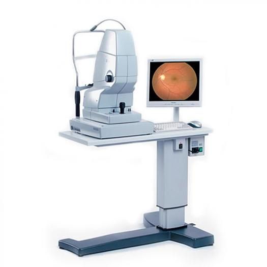
ZEISS Visucam ProNM FA, FAF, ICG
AMERICA North (USA-Canada-Mexico)
The Zeiss VISUCAM PRO NM Non-Mydriatic Fundus Camera increases the quality
and simplicity of fundus imaging. Compact, yet big enough to set the standard in
ophthalmic photography, The Zeiss VISUCAM PRO NM features a unique combination
of functions to enhance fundus visualization and documentation.
The Zeiss VISUCAM PRO NM is designed for both routine clinical use and screening.
The Zeiss VISUCAM PRO NM integrates all elements of clinical retinal photography –
from image capture to image documentation – in a single, state-of-the-art system
featuring all hardware and software.
Operation is easy to ensure a smooth, rapid workflow with the help of
the positioning aid with working distance dots, a focusing aid with
paired coincidence lines and ergonomic design.
When using the Zeiss VISUCAM PRO NM the visual overview and assessment are
possible at all times in every phase of the exam. When the image is
captured on the Zeiss VISUCAM PRO NM, it immediately appears on
the 17′′ flat screen monitor and is automatically stored.
With the Zeiss VISUCAM PRO NM’s 3D images and 45° and 30° field angles,
the excellent image quality of the Zeiss VISUCAM NM makes it the perfect solution
for cases which require in-depth study. Software manages image display,
editing, printing and data export. A variety of image export formats are available
with the Zeiss VISUCAM PRO NM.
Zeiss VISUCAM PRO NM Fundus Camera Features:
The following capture modes are available with the VISUCAM NM/FA, FAF, ICG:
The intuitive, easy-to-use software combine positioning and focusing aids into a smooth
and fast workflow. Each task is quickly performed increasing your workflow.
The stereo module provides a convenient capability for taking and
displaying 3D images. ZEISS AutoMap automatically generates montages of
large areas of the retina from peripheral images, and, with the separation of green,
blue and red color channels from existing color photos, fewer exposures are necessary.
Essential elements for a fast workflow include intuitive internal fixation and the
Autoflash mode – you concentrate on the patient. Existing and new image data can
be analyzed at any time at the same workstation, and compared or
easily forwarded through your network on a USB stick or with the included DVD writer.