 1/1
1/1
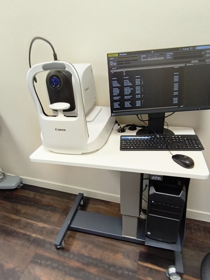
Canon Xephilio OCT-A1
EUROPE (Western and Northern)
Pre-owned equipment inspected by our experts
Functional condition: Very good
Cosmetic condition: Very good
Canon reliability and precision
Supplied with table and PC
Technical specifications
Xephilio OCT-A1: Canon Medical’s latest high-resolution (3 μm)
optical coherence tomography system, delivering remarkable
performance and exceptional ease of use for your daily practice.
High-definition image quality and a host of automated
features optimize and simplify examinations,
ensuring fast acquisition times while boosting efficiency
and patient comfort.
High-resolution OCT
The Xephilio OCT-A1 delivers exceptional image quality.
An optical resolution of 3 microns (1.6 μm digital resolution)
enables excellent differentiation of retinal structures and
individual layers.
Complete examination in just 3 clicks
Examinations with the Xephilio OCT-A1 are extremely straightforward,
making them easy to delegate. Obtaining scans requires just
three mouse clicks. A comprehensive suite of intelligent
features enables fully automated examinations.
Additionally, an automatic re-scan function activates if
the patient makes unwanted eye movements,
automatically compensating for motion artifacts.
Automated anterior segment tracking
After a single mouse click near the center of the pupil,
the OCT-A1’s automated anterior tracking system takes over,
automatically detecting and maintaining the precise center—even
if the patient moves their eyes or blinks.
Automatic image optimization
A second mouse click initiates the scan, and the device
automatically optimizes focus and the coherence gate to
ensure the best possible signal quality and examination results.
After confirming the preview scan images, a third click launches
the actual acquisition, which takes only a few seconds.
Real-time retinal tracking
The OCT-A1 is equipped with a scanning laser ophthalmoscope.
This SLO system enables real-time retinal tracking. By detecting
and compensating for movement, the impact of
small involuntary motions—which could otherwise cause
motion artifacts—is significantly reduced,
greatly enhancing image quality.
High image resolution
With the Xephilio OCT-A1, up to 200 scans can be averaged to
achieve an image resolution that reveals the detailed structure
of the retinal layers as well as the folded structure of the vitreous.
The device combines high image resolution with
an exceptionally wide scan range of up to 13 mm.
Enhanced depth visualization
For optimal imaging, the system offers specialized scan modes
for vitreous and choroidal imaging.
Reliable 10-layer recognition
Canon’s Xephilio OCT-A1 can automatically detect and
distinguish 10 retinal layers, including Bruch's membrane (BM).
Anterior segment imaging
With the optional ASA-1 anterior segment adapter,
the Xephilio OCT-A1’s 3-micron resolution also allows for
high-resolution imaging of the eye's anterior segment.
Anterior scans can be performed with a width of 6 mm or 9 mm.