 1/1
1/1
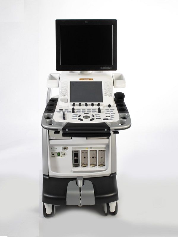
GE Vivid E9 XDclear
EUROPE (Western and Northern)
GE VIVID E9 XDclear – Refurbished Ultrasound
Extraordinary capabilities
Whether in TTE or TEE, capture the entire heart in a single beat. Image a valve,
or the entire ventricle with excellent image quality. And break down the barriers to
routine day-to-day 4D imaging, from acquisition, through navigation and quantification,
to archiving.Vivid E9 is GE Healthcare’s first cardiovascular ultrasound system built
specifically for 4D imaging?from ergonomics to image acquisition to data management.
Available in both 2D and 4D configurations.
Featuring our Accelerated Volume Architecture 4D platform for
increased processing power compared to GE Healthcare’s previous 4D scanner.
Advanced quantification tools help streamline workflow for enhanced productivity.
These tools include 4D Stress and 4D Auto LV Quantification. One-touch ease of use and
intuitive workflow puts 4D at your fingertips. As 4D becomes routine in
your clinical practice, you still need excellent 2D image quality.
Vivid E9 handles 2D imaging with equal power, precision, and agility,
made possible with our 4V-D transducer, the secondgeneration 4D transducer,
and the M5S/M5Sc transducers single-crystal matrix array and
XDclear transducer technology. Open the door to 4D and its many new possibilities.
Accelerated Volume Architecture GE’s exclusive and patented beam-forming technology,
the Accelerated Volume Architecture, provides several times the power of
traditional GE ultrasound systems with increased volume size for full volume single beat,
as well as for highvolume rate multi-beat 4D acquisition. Using both coherent and
harmonic image processing, the system provides computational power, ease of imaging,
workflow flexibility and product upgradeability.
Raw data format The GE Vivid product line has always acquired and stored its data in
a specific raw data format, which enables onboard, as well as after-the-fact post-
processing capabilities. This flexible and innovative format and storage of
pre-scan converted data has enabled development of utilities with high clinical value.
This has resulted in an increase in the ability to perform additional advanced
algorithms for all of the steps in the data processing chain, culminating in
the release of the Vivid E9 with XDclear.
Extraordinary image quality
Crisp imaging. Focused workflow. Clear quantification. Vivid E9 makes short work of
your routine 2D exams.
2D image quality Designed with GE’s proprietary XDclear technology,
the M5Sc transducer delivers excellent endocardial definition and texture.
Together with controls like UD Clarity and HD imaging, it helps provide crisp valves and
borders across a wide range of patients. Similar image quality is achieved with
pediatrics, vascular, abdominal, and TEE transducers.
Extraordinary quantification
Based on extensive feedback from clinicians just like you, TTE and TEE on the Vivid E9
are all about making imaging simple, intuitive, and quantifiable to
help make your work easy and efficient.
Automated Function Imaging (AFI) This software tool assesses and
quantifies left ventricular wall motion at rest. It calculates a large set of parameters to
describe the function of the left ventricular walls.
AFI specifically calculates peak systolic longitudinal strain (both segmental and global)
and presents the results as parametric images.
As part of its healthymagination validation, a study has shown that AFI offers potential in
predicting mortality in patients with suspected LV impairment compared to
Ejection Fraction and Wall Motion
Extraordinary workflow
The Vivid E9 helps make 4D imaging as easy as 2D imaging.
It can bring remarkable enhancements to your entire 4D workflow process,
thanks to fast, consistent reproducibility.
4D Views
4D Virtual Store
Advanced 4D User Tool Box
4D Stress
Scan Assist
Scan Assist Pro
Extraordinary ergonomics
From its slim, lightweight, maneuverable design, to its adjustable electronic keyboard,
the Vivid E9 system is ergonomically designed to be easy to handle and operate.
Adjustable LCD display
Highly mobile
User adaptable
Front and rear handles
Accessible touch panel controls
Adjustable floating keyboard
Easy keyboard storage
Convenient data management
ADDITIONAL INFORMATION
WEIGHT 140 kg
DIMENSIONS 55 × 85 × 115 cm
MEDICAL SPECIALTY Cardiology
COLOR Color Flow Mode, PowerFlow
DOPPLER CW Color Doppler, PW Color Doppler, Real-time Doppler Auto Trace
DISPLAY TECHNOLOGY LCD Monitor
IMAGE QUALITY High Definition, SXGA (1280 x 1024)
SCREEN DIMENSIONS 17" inches
CONNECTIVITY DICOM, DVI-D, Ethernet, USB 2.0, DVD External
PRINT TYPE Ethernet Color Printer, Thermal B/W Printer, USB Color Printer
HEAD PORTS Electronic scanning probes: 4 active
SYSTEM PORTABILITY Wheeled
Compatible Probes
Convex
C1-5-D
4C-D
C2-9-D
8C
Endocavity
IC5-9-D
Linear
9L-D
11L-D
ML6-15-D
i13L
Phased Array
M5S-D
M5Sc-D
12S-D
6S-D
Matrix 3D-4D
4V-D
Independent CW Doppler
P2D
P6D
TEE Cardio
6VT-D
6Tc
9T