 1/1
1/1
GE
AMERICA North (USA-Canada-Mexico)
Features
Speckle Reduction Imaging (SRI) heightens your visibility of organs and lesions with high-definition contrast resolution that suppresses speckle artifact while maintaining true tissue architecture.
CrossXBeam enhances tissue and border differentiation with real-time spatial compounding acquisition and processing.
B-Flow Color allows you to see hemodynamics in great detail to distinguish more easily between different flow variations, while maintaining high frame rates. Achieve exceptional temporal and spatial resolution when scanning vessels by directly imaging blood reflectors.
Raw Data allows data to be stored early in the image chain for optimum flexibility during post-processing and analysis.
Single-crystal transducers use new ceramic materials to increase bandwidth and improve sensitivity to deliver exceptional cardiac performance, even for your most difficult patients. Obtain crystal-clear images in every exam regardless of patient size.
Portable and powerful enough to go almost anywhere, and give you the exceptional imaging you need.
Real-time 4D imaging provides the power to acquire and construct volumetric images, displaying multi-planar anatomical views not otherwise visualized.
DualBeam maintains high frame rates while using high line density, even in abnormal settings. Increase temporal resolution in fast-flow cardiac and vascular studies.
Harmonics increase resolution and cystic clarity with a combination of coded harmonics and Phase Inversion Harmonics.
System Specifications
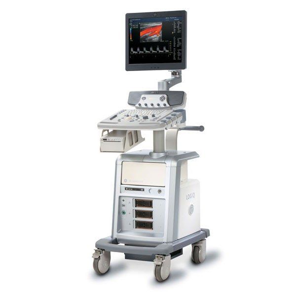
Height: 1360 mm
Width: 430 mm
Depth: 640 mm
Weight: approx. 80 kg (176 lb.)
Monitor: 17-inch TFT LCD
Applications
Abdominal
Obstetrical
Gynecological
Cardiac
Musculoskeletal
Vascular
Urological
Small Parts and Superficial
Breast
Pediatric and Neonatal
Transcranial
Endocavity
Intraoperative
Transesophageal
Transducer Types
Sector Phased Array
Convex Array
Microconvex Array
Linear Array
Single CW (Pencil)
TEE Multiplane Sector Array
Bi-plane Microconvex Array
Volume ‘4D’
Operating Modes
B-Mode
Coded Harmonic Imaging
M-Mode
Color Flow Mode (CFM)
Power Doppler Imaging (PDI) with Directional Map
PW Doppler w/High PRF
M-Color Flow Mode
Anatomical M-Mode (option)
Anatomical M-Color Mode (option)
B-Flow Mode (option)
B-Flow Color Mode (option)
Coded Contrast Imaging (option)
CW Doppler Mode (option)
PFD Mode (option)
Tissue Velocity Imaging (TVI) Mode (option)
3D/4D Volume Modes (option)
3D Static (option)
4D Real time (option)
Elastography (option)
Transducers
GE 4C – 1.4-5.0MHz, Convex
GE 5CS – 2-6MHz, Convex
GE 8C – 4-11MHz, Microconvex
GE E8C – 4-11MHz, Microconvex (Endocavity)
GE E8CS – 4-11MHz, Microconvex
GE 3CRF – 2-4MHz, Microconvex
GE BE9CS – 4-11MHz, Bi-plane Microconvex
GE 8L – 4-11MHz, Linear
GE 9L – 3-10MHz, Linear
GE 11L – 5-13MHz, Linear
GE ML6-15 – 6-15MHz, Linear
GE T739 – 4-12MHz, Linear (Intraoperative)
GE i739 – 4-12MHz, Linear (Intraoperative)
GE i12L – 5-12MHz, Linear (Intraoperative)
GE 3Sp – 1.5-5.5MHz, Phased Array Sector
GE 5Sp – 4-10MHz, Phased Array Sector
GE 3S – 1.5-3.5MHz, Phased Array Sector
GE 5S – 2-5MHz, Phased Array Sector
GE 7S – 3-8MHz, Phased Array Sector
GR 6Tc – 3-7MHz, Multiplane Transesophageal Phased Array Sector
GE ERB – 3-11MHz, Bi-plane Intracavity
GE 4D3C-L – 2-5MHz, Convex Volume
GE 4D5C-L – 3-7MHz, Convex Volume
GE 4DE7C – 4-11MHz, Convex Volume
GE 4D8C – 4-11MHz, Microconvex Volume
GE P2D – 2.0MHz, Non-Imaging Single CW Doppler Pencil
GE P6D – 5.0MHz, Non-Imaging Single CW Doppler Pencil