 1/1
1/1
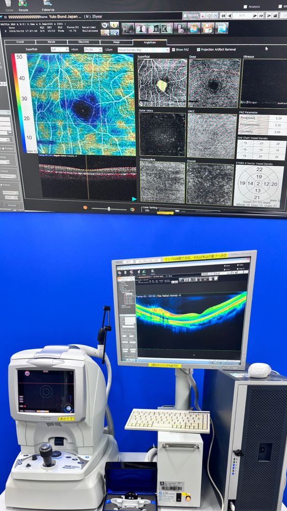
Nidek RS-3000 Advance
ASIA (North East)
Nidek OCT RS-3000 Advance comprised of the main body for
capturing images and a storing with computer.
OCT Angiography is a non-invasive technique that does not require injection
of contrast dye for layer-by-layer examination of the retinochoroidal vasculature.
OCT Angiography allows greater visualization of deep and small capillary plexus which
is a significant advancement for clinical evaluation of retinal disease compared
to conventional OCT or fluorescein angiography.
Nidek OCT RS-3000 Advance with ANGIOGRAPHY OCT system
incorporating Scanning Laser Ophthalmoscope (SLO) designed
to an evaluation choroid and the retina.
Wide Area Scan 9 x 9
Specification & Options
This RS-3000 Advance model is upgraded to AngioScan OCT by
a simple software upgrade. The functionality of RS-3000 Advance combined
with this software allows evaluation of retina in greater detail helping with
the diagnosis and enhancing patient care.
Key Features
-Retinal Camera and OCT combined
-Anterior imaging for pachymetry
-Anterior imaging for angle view
-Superb macula image using SLO technology
-Wide-field imaging, from macula to disc
-Tracing HD plus for accurate
-Selectable OCT sensitivity enables enhanced visualisation
-Accurate image capture using Torsion Eye Tracer (TET)
-Multifunctional follow-up Customised report
-Angiography Option is included.
Configuration
Main OCT Unit, a key board, PC, Monitor,
attachment for anterior, anterior lens kit for RS-3000 advance
Scan speed: Max. 53,000 A-scans / s
Power Supply : AC 100, 120, 230 V 50 / 60 Hz
Dimension : 380 (W) x 524 (D) x 499 to 531 (H) mm / 34 kg
15.0 (W) x 20.6 (D) x 19.6 to 20.9 (H)“ / 75 lbs
Condition
Patient Ready