 1/1
1/1
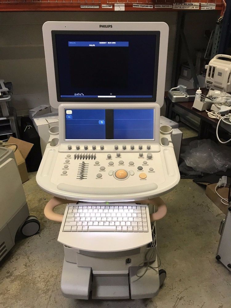
Philips iE33
ASIA (South East)
ULTRASOUND SYSTEM WITH X3-1, S5-1 & L8-4 TRANSDUCERS
THE PHILIPS IE33 ULTRASOUND SYSTEM IS
A PREMIUM LEVEL CARDIOVASCULAR ULTRASOUND MACHINE
THAT WAS LAUNCHED IN 2004
AND HAS BEEN REVISED AND IMPROVED OVER THE PAST DECADE.
Live 3D:
– x MATRIX cardiac transducer with 3000 elements
– Automated Stress Echo
– Premium Image Quality
– Full Doppler functions
– Live 3D TEE
– 20-inch high resolution articulating flat panel display
– Fully articulating control panel
– Live X plane multi plane imaging
– SonoCT Compound Imaging
– Tissue Harmonics and Tissue Harmonic Sono CT
– X RES
– i SCAN one-touch optimization
– i FOCUS one-button focal zone optimization
– i OPTIMIZE one-button optimization multifunction auto image optimization
Integrated CD/DVD burner
– Export to AVI and JPG
– DICOM 3.0
– USB port for import/export of images
– On-Board workstation data management
– 8-speaker high-fidelity stereo audio
– Panoramic Imaging
– Channels scalable up to 57,000 channels
– Multiple triplex mode operation
– High Resolution interactive color touch panel with ambiance adjustments
– Pull-out alphanumeric keyboard
– 2D/M-Mode
– Color Power Angio
– CW/PW/Color Doppler
– Duplex CW Doppler
– Cine Loop
– User-Defined presets and calculations
Applications & Usage
– Clear views of cardiac anatomy for the most accurate EF calculations
– Live 3D volumes provide high frame rate and high image quality
– More clinical information available in one view with Live #D and other tools
– Utilizes the next generation of SmartExam for echo studies
– Auto Doppler helps with image and sample volume placement
Advanced Feature Details
– Choose 2D, 3D or a combination without disrupting workflow
– Highly ergonomic X5-1 transducer
– PureWave Transducer Technology allows for imaging for a wind range of patients
– Provides fewer artifacts, better penetration than conventional transducer technology
– Advanced XRES completed 350 million calculations per frame
– Adaptive Broadband flow automatically adapts the frequency to ROI to enhance spatial resolution
– Access data from PACS on the iE33
– Simple way to compare past and current studies without the use of a reading station
Speed Up Echo Lab Workflow
– Add 3D into any exam at any time
– Gather more accurate LV volume measurements
– Calibrated measurements on the Live 3D volume or MPR views without a quantification program
– Image whole heart in 3D and real time in one cardiac cycle
– iCrop easily focuses on structures within a volume
– Live Full Volume mode offers functionality to capture a whole left ventricle and eliminate foreshortening
– Visualize subtlve valve leaflets in 3D and 2D
– Utilize 3DQ Advanced to generate a full 3D endocardial border
Additional Workflow Tools
– Improved 2D EF calculations with LivexPlane
– Obtain realtime images of the entire hear using Live Full Volume
– Zoom in on one volume region of interest with iCrop
– Utilize semi-automaticed, on cart and off cart analysis with 3DQ Advanced
– Cardiac Motion Quantification (CMQ) allows for assessment of global and regional cardiac function
Stress Echo Exam Capabilities
– Q-station software allows for easy viewing, quantifying and reporting on any PC
– Easy integration of stress echo and ECG ST elevation maps into a single report
– X5-1 transducer provides high image quality in both 2D and 3D modes
– iRotate Stress allows for completion of the entire stress echo protocol without rotating the transducer
– Cardiac Motion Quantification (CMQ-Stress) allows for quantifying 2D stress echo settings
– iSlice allows for �slicing� of views to find the best content
– Pre-loaded 2D/3D Stress protocol
Pediatric Echocardiography
– Complete transeophageal echo on patients less than 3.5 kg
– Perform TDI strain analysis on smaller patients
– Live 3D zoom and 3D color allows for easy access to assess flow defects
– Live xPlane allows users to pull two simultaneous orthogonal views without rotating the transducer
Remote Service Tools
– Remote desktop allows Philips service engineer to get a live view of your system�s console for real-time troubleshooting and faster issue resolution
– iSSL Technology meets global privacy standards and provides a safe and secure connection when connecting to Philips remote services
– Place a technical issues request directly with Philips without interrupting workflow
– Proactive support from Philips helps users detect issues before can impact performance.
SOFTWARE VERSION: 3.0.2.711
CART LEVEL: A
MFG: 2005
APPLICATIONS:
– Cardiac
– Vascular
INSTALLED OPTIONS:
– 2DQ
– 3DQ Advanced
– 3DQ Basic Measurement/Quantification
– Acquisition Protocol (Stress Echo)
– Netlink DICOM 3.0
– IMT
– Live 3D
– ROI Tools
– Strain Q
– Tissue Doppler Imaging
– iCommand
– Language Option: English
– Clinical Option: Cardiac & Vascular
PHILIPS iE33 ULTRASOUND SYSTEM ACCESSORIES:
– Mitsubishi MD3000 VCR
PHILIPS iE33 ULTRASOUND SYSTEM TRANSDUCERS
– X3-1 3D/4D xMatrix Array
– S5-1 Sector Array
– L8-4 Linear Array